Sample Preparation
Homogenization
Heating and Mixing
Electrophoresis and Blotting
Polyacrylamide Gel Electrophoresis
Agarose Gel Electrophoresis
Western Blotting
Power Supplies
PCR & qPCR Thermal Cycler
Thermal Cycler (PCR)
Real-time Thermal Cycler (qPCR)
PCR Workstations & Cabinets
UVP BioImaging Systems
Molecular Spectroscopy
Lab Equipment
Ultraviolet Products
Hybridization Ovens
UVP Incubator
UV Crosslinkers
UVP Benchtop Transilluminators
Thermal Mixers
Electrophoresis & Blotting
Thermostats
View All
Fume hood
Laminar Airflow
Biosafety Cabinet
Autoclave
Centrifuge
pH Meter
Shaker & Mixer
Orbital Shaking Incubator
BOD Incubator
Heating Oven
Water Purification System
Aermax - Air Purification
Medical Oxygen Concetrators
Hygiene Solution
-150°C Cryogenic Freezer
-86°C Ultra Low Temp Freezer
-40°C Low Temp Freezer
-18 ~ -25°C Biomedical Freezer
-20°C Biomedical Freezer
4° ± 1°C Blood Bank Refrigerators
2~8°C Pharma Refrigerators
2~8°C ICE Lined Refrigerators
-25°C ~ + 4°C Mobile Freezer/Collers
20~24°C Blood Platelet Incubators
Ice Machines
Coldrooms
Mortuary Chambers
Microscopy techniques have revolutionized pathological studies by their great detailed study and observations. Almost 200 years ago, the microscopy technique came to light which has eased out the pathological studies and the research observations. In recent times, molecular testing techniques are pacing at a good speed, as they are studied for getting insights of the concerned biological specimen under a trinocular compound microscope through the trinocular head. For imaging of all types of cells and tissues, the pathologists need an intelligent microscope that would help in the magnification of inconspicuous cells and tissues, and also help in making detailed observations of it. These microscopes are all called culture microscopes, cell culture microscope, and tissue culture microscope. Some advanced microscopes use the digital method for observing the cells and samples. This microscopic technique uses the camera for recording the bioimaging of all layers of the biological specimens.
In pathological studies, various microscopic techniques are used which are used to extract the details from the cell cultures and biological samples.
Before the invention of microscopes, pathological studies were performed by using some simple techniques which could not yield good results. There were no insights extracted from the study of cells and samples by using manual methods. To extract every small detail out of the cell cultures and biological specimens in a pathology lab, a microscope is needed. With the help of a microscope, pathologists can easily view the magnified images of all types of cells and tissues.
Some microscopic techniques, like phase contrast microscopy and darkfield microscopy technique, are used for illuminating the images. So, the images can be easily deciphered by the pathologists for any type of anomaly. On the contrary, the simple and compound microscope uses the simple microscopy technique to magnify the images of all types of cellular structures.
In digital pathology techniques, the best trinocular microscope is fitted with good resolution cameras for recording the images. In addition to all these, a computer system is also fitted to the system for providing a more detailed overview of the specimens.
Following are the various types of microscopes used in pathological studies :
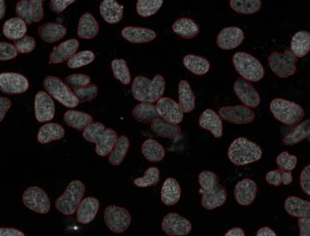
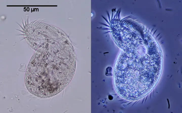
Phase contrast microscopic devices are also called tissue culture microscopes, because of their contrasting ability to illuminate the image. When the direct source of light is made to strike in the biological sample, the light starts to get diffract. This diffraction of light produces the interference phases which are not that subtle to be seen with naked eyes. While scattering the light or diffraction of light, some of the light gets absorbed by the specimen illuminating it with full contrast. However, the light which is not diffracted in the phase contrast microscopy is called zero-order light. Here, the zero-order light remains unmodified throughout the magnification process of bioimaging. The pathologist makes use of this microscopic technique to visualize the microorganisms which are too difficult to be seen from the naked eye.
This microscopy technique observes or visualizes a living organism in its natural state or its native environment. It can provide far more information than specimens that need to be killed, fixed, or stained to view under a standard compound microscope.
The phase contrast microscope gives out high-contrast, high-resolution images of all biological samples or specimens.
The inverted microscope is similar to light microscopes which give out better images of biological specimens. So, they are also used in the observations of cell culture and tissue cultures. So, it is called a cell culture microscope. Like the phase contrast device, the inverted microscope with the camera also gives out detailed and structured images. This makes the inverted microscopy technique one of the good options to view the specimens. The images formed in the inverted microscope are inverted, i.e, the visualization is done from the bottom to top.
The inverted phase contrast microscopes use the working mechanism of phase contrast microscopy to give out the high contrast images in an inverted manner. Interferences are also used in the magnification process of biological cell cultures in pathology labs. Apart from all these things, several modern devices, like cameras and computer systems can also be used. The devices like cameras, illumination devices, fluorescence devices, computer networks, and many other image capturing devices can be fitted to the inverted phase contrast microscope. In addition to this, confocal scanning devices can also be used to get a better-magnified image of biological cells and cultures.
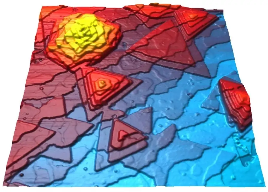
The light source in the darkfield microscope showers the light on the sample which gets scattered by the special condensers fitted into the microscope. As the light rays move past the biological culture or cell plane towards the apex of the hollow cone. The light rays pass on the move to the hollow cones prepared by the reflected light. Here, the light rays travel past the objective lens in the darkfield microscopic device, which seals the entry of other light rays in the trinocular compound microscope. The entire observational area appears dark when there is no biological specimen on the stage. When a biological sample is placed on the pointing stage, the light apex cone strikes it and generates the image. Hence, this microscopy technique is named a darkfield microscope. All the light rays scatter through the condensers are captured by the objective lens which further generates an image of the biological specimen. The image formed here is highly illuminated against the darker background to make the cells more visible. This image illumination technique in a darkfield microscope makes it favorable to the use of pathologists.
Trinocular Compound microscopes are made up of two lenses which offer better magnification than the traditional simple microscope. In this case, the second lens magnifies the images of the first lens. These compound microscopes are brightfield microscopes in which the specimen is lit underneath the pointing stage. The images formed in the trinocular compound microscope can be binocular or monocular.
The trinocular compound microscopes provide a magnification of 1,000 times more than the simple microscopes. But the resolution of the image formed is low, because of this, the compound microscopes are not that much in use. However, the trinocular compound microscope allows the pathologists to take a closer look at the cells which are too small to be seen by the naked eye. Individual cells or aggregate of cells can also be magnified by the use of a compound microscope. The specimens used in the compound microscope’s magnification are relatively transparent than the other biological specimens. In a manner of cost-effectiveness, the compound microscope is a little more expensive than the simple microscopes. Because of its high power of magnification, it is used in biology labs and pathology for studying cultures and individual cells.
Simple microscopes are generally considered the first microscope to be used for the observations. It was created in the 17th century by Anton van Leeuwenhoek, by combining a convex lens with a holder for the specimens used. The magnifying power of this simple microscope is between 200 and 300 times which is good for the magnification of the biological specimens. The red blood cells were the first microscopic cells which are studied with a simple microscope. Today, simple and traditional microscopes are not that much used because of their low magnification power. Despite all these, the simple microscope is the base of all advanced microscopy techniques which we use in our daily biology experiments. This has led to the discovery of many powerful compound microscopes in the scientific world. Some transparent tissues or cells are well magnified in the simple microscope.
Conclusion
The pathologists make use of all the above-mentioned microscopes in the study of the cells and other samples. The rise of microscopes has led to proper diagnostics of the diseases caused by the pathogens present in the biological specimens.