Sample Preparation
Homogenization
Heating and Mixing
Electrophoresis and Blotting
Polyacrylamide Gel Electrophoresis
Agarose Gel Electrophoresis
Western Blotting
Power Supplies
PCR & qPCR Thermal Cycler
Thermal Cycler (PCR)
Real-time Thermal Cycler (qPCR)
PCR Workstations & Cabinets
UVP BioImaging Systems
Molecular Spectroscopy
Lab Equipment
Ultraviolet Products
Hybridization Ovens
UVP Incubator
UV Crosslinkers
UVP Benchtop Transilluminators
Thermal Mixers
Electrophoresis & Blotting
Thermostats
View All
Fume hood
Laminar Airflow
Biosafety Cabinet
Autoclave
Centrifuge
pH Meter
Shaker & Mixer
Orbital Shaking Incubator
BOD Incubator
Heating Oven
Water Purification System
Aermax - Air Purification
Medical Oxygen Concetrators
Hygiene Solution
-150°C Cryogenic Freezer
-86°C Ultra Low Temp Freezer
-40°C Low Temp Freezer
-18 ~ -25°C Biomedical Freezer
-20°C Biomedical Freezer
4° ± 1°C Blood Bank Refrigerators
2~8°C Pharma Refrigerators
2~8°C ICE Lined Refrigerators
-25°C ~ + 4°C Mobile Freezer/Collers
20~24°C Blood Platelet Incubators
Ice Machines
Coldrooms
Mortuary Chambers
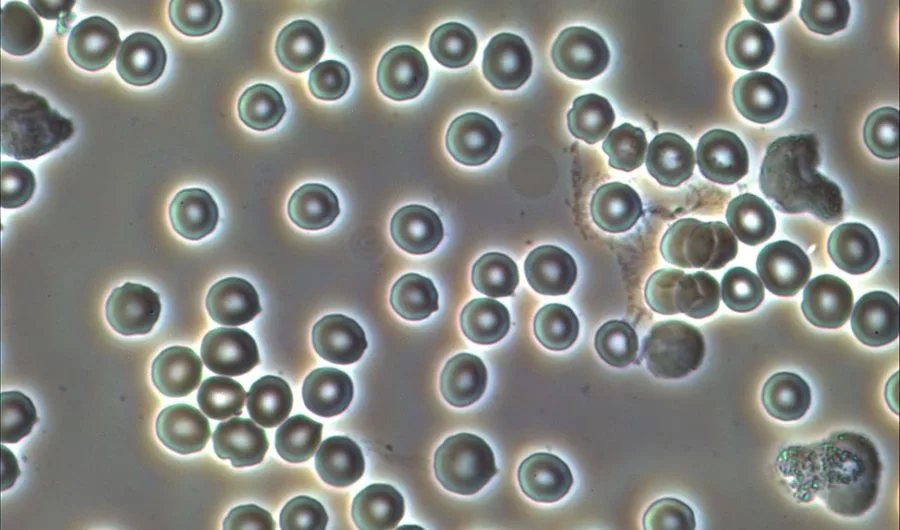
Since the discovery of phase contrast microscopy, it is used widely for several observations over biological specimens. In the year 1934, Dutch physicist Frits Zernike discovered it by modifying the components of a traditional trinocular compound microscope. This microscope works on the principle of phase differences caused by the interference processes. The phase-contrast microscope is worth converting all phase differences into amplitude variations which can be easily detected by the naked eyes also. In the scientific world, the inverted phase microscope is used for the production of high-contrasted images of all types of transparent specimens like living epithelial cells and others. The main cause behind the production of contrasted images is the interference process that requires partial coherent light to illuminate the biological sample. This source of light should be longitudinally coherent and precise to make it work with phase contrast microscopy.
Generally, in a phase contrast microscope, the numerical aperture (NA) of the concerned condenser is reduced by the geometrical arrangement of annular rings. This reduction in the condenser helps in the illumination of the transparent specimens under a trinocular head. The reflection and diffraction of light rays happen in the microscope for the production of high contrasted images of all samples. It mainly changes the phases of the diffracted light beam instead of the amplitude of the concerned light beams. Illumination is easily visible to the naked eyes of researchers and scientists while observing the sample on the pointing stage.
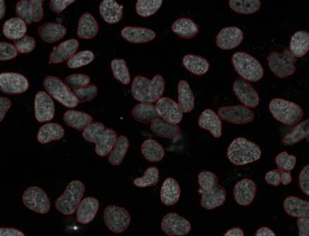
Zernike’s discovery of phase contrast microscopy has led to many critical observations in the biological world. Because of this, the study of all types of transparent specimens can be done easily and efficiently. Due to the inverted phase contrast microscope‘s ability to enhance the contrast of transparent, unlabelled specimens, it is used widely for the observations of living cells or cell imaging. For transparent specimens, the illumination of light rays makes up the high contrasted images, but for the small culture and multi–wells, the beam of illuminated light gets distorted. This happens due to the presence of buffer solutions, the light beams get curved over the surface of the liquid which further distorts the illumination path light. All these lead to a misalignment of ring-shaped illumination concerning the phase rings of the phase contrast microscope. Here, the phase contrast microscope’s phase rings are present behind the objective lens. As a consequence of this, the images formed contrast and quality can get severely damaged. It happens especially when the imaging is done at the margins of the containers.
For getting a perfect illumination, the process of adjustment of phase rings should be done properly. To avoid misalignment of the light rays, the observations should be done through transparent specimens, instead of using containers. The microscopes which are used for cell culture and tissue culture are called cell culture microscopes, culture microscope, and tissue culture microscope.
For demonstrating the practical implementation of phase contrast, the following components are required :
A light source is used in the magnification process of phase contrast microscopy. Two beams of light rays get emitted by the light source, get focussed by the field lens exactly inside the opening of the condenser annular ring. As this location is precisely in the front of the focal plane of the condenser, the two light rays start to refract. While refracting process, the light rays exit the condenser as parallel rays of light. It is assumed that the two light rays are neither refracted nor diffracted in the specimen plane. These light rays enter the objective lens of the inverted microscope as parallel beams of light.
It is seen that all parallel rays get focussed in the back focal plane of the objective. Here, the back focal plane is a conjugated aperture plane to the condenser’s front focal plane. This focal plane is located at the condenser annulus. For completing the phase setup of the concerned microscope, a phase plate is positioned inside the back focal plane in a way that it gets lined up nicely with a condenser annulus. The location of the phase plate is present in the suitable location inside the objective lens. And the annular planes are present in the conjugated aperture planes which are perpendicular to the optical axis.
To get the proper illumination of the biological specimens, the centering of two elements is done very precisely. By this method, the phase contrast illumination can be established. A phase centering telescope can also be used in the upright and inverted microscope to replace one of the oculars used to center the annular ring with the ring of the phase plate of the microscope.
The light rays first reach the condenser annulus through a small aperture located in the center. The phase plate is also just covering a small aperture located in the plane labeled area. All light rays diverge through this annulus of the condenser and the phase plate is also covering a small aperture. In the phase contrast microscopy, the optical system is greatly simplified by showing only two single lenses that represent the optical elements. After the mediation of light rays, the microscope tends to start illuminating the images by using the phase differences. All biological specimens start to get illuminated by the process of phase differences. Some of the light beams remain un-interfered which are discarded out of the phase contrast microscope. The best trinocular microscope also has this feature of illumination that helps in observations of the biological specimens.
In Zernike’s phase contrast microscopy, the image formation is strongly based on the central phase contrast treated images. In this microscopy technique, the cone-shaped illumination; geometry ensures the magnification of the biological specimens. This increases the resolution of the images and the axial sectioning. The precondition of a perfect match between the illumination and filter also ensures perfect illumination. In multi-well cultures, small – small filters are present which are made up of a single polymer holder. These microwells are used ubiquitously in biomedical research and clinical settings. This allows the cells to get exposed to a variety of different conditions to study the effects. With a perfect illumination cone, the biological specimens can be studied efficiently.
Conclusion
Phase contrast microscopy techniques are useful in observing the living cells in a proper structural way. Because of its illumination cone and diffraction and interferences, the images can be studied with sheer observations. You can check the price of the trinocular microscope online as well as an offline medium for your laboratory. Illumination is the consequence of different phase distributions in the source light rays. The condenser lens and objective lens are used to mediate the light rays from the light source to the surface of the biological specimen.