Sample Preparation
Homogenization
Heating and Mixing
Electrophoresis and Blotting
Polyacrylamide Gel Electrophoresis
Agarose Gel Electrophoresis
Western Blotting
Power Supplies
PCR & qPCR Thermal Cycler
Thermal Cycler (PCR)
Real-time Thermal Cycler (qPCR)
PCR Workstations & Cabinets
UVP BioImaging Systems
Molecular Spectroscopy
Lab Equipment
Ultraviolet Products
Hybridization Ovens
UVP Incubator
UV Crosslinkers
UVP Benchtop Transilluminators
Thermal Mixers
Electrophoresis & Blotting
Thermostats
View All
Fume hood
Laminar Airflow
Biosafety Cabinet
Autoclave
Centrifuge
pH Meter
Shaker & Mixer
Orbital Shaking Incubator
BOD Incubator
Heating Oven
Water Purification System
Aermax - Air Purification
Medical Oxygen Concetrators
Hygiene Solution
-150°C Cryogenic Freezer
-86°C Ultra Low Temp Freezer
-40°C Low Temp Freezer
-18 ~ -25°C Biomedical Freezer
-20°C Biomedical Freezer
4° ± 1°C Blood Bank Refrigerators
2~8°C Pharma Refrigerators
2~8°C ICE Lined Refrigerators
-25°C ~ + 4°C Mobile Freezer/Collers
20~24°C Blood Platelet Incubators
Ice Machines
Coldrooms
Mortuary Chambers
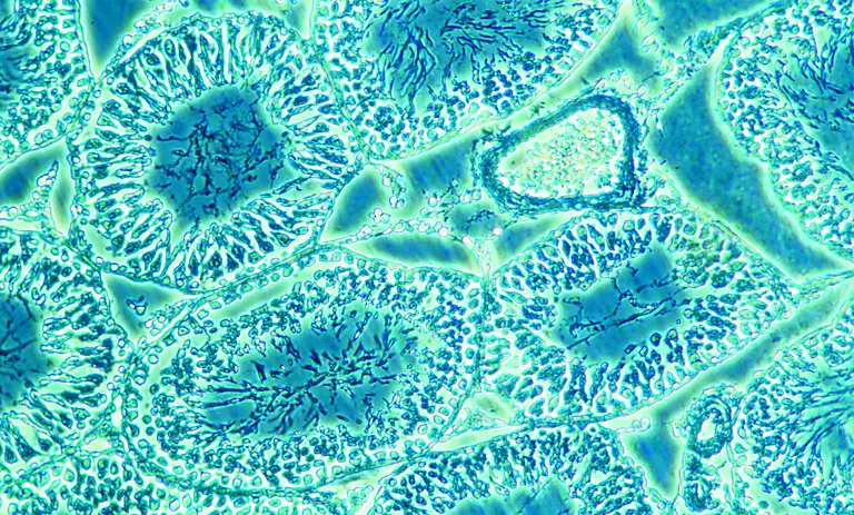
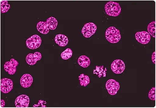
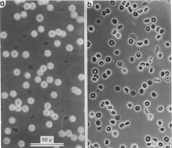
Phase-contrast microscopy comes in the category of light microscopy which offers a powerful technique of imaging biological samples. The cells are visualized under the microscope in label-free imaging solutions. Phase contrast microscopes use the mechanism of light microscopy for making the vision of cells in a good way. Label-free imaging of biological samples is done for understanding more cell structure, functions, and behavior. The microscopy techniques are used to get the enlarged and highly magnified image of cells. Traditional or standard light microscopy has the limits of setting a level of contrast. This makes the magnification not clear. As the cells don’t absorb light and also reflect the light which makes a change in their refractive index. However, the refractive index also scatters the light which reveals the structure of the cells. This makes the cell’s magnification a little blurry. But when you use a phase-contrast microscope, you overcome all these problems in the cell’s magnification.
With light microscopy, you cannot get the required image of the biological samples. This makes a problem for label-free imaging of the biomolecules and other research applications. On the other hand, a researcher or scientist can use the inverted phase-contrast microscope, they can get the required imaging of biological samples. The transparent specimens like cells are placed under a tissue culture microscope within a required contrast level to get the magnified sample of the image. In a phase-contrast microscope, it converts the different optical wavelengths or the different refractive index of the biological samples into shifts in the direction of the phase of light. These shifts of light are used to create a final contrast. This makes the biological samples more magnified and thus a trinocular microscope of phase-contrast nature can be used. It can give the researchers the label-free imaging of various biological samples.
In a trinocular head microscope, the light phase is present in a mathematically described sine wave. The light phase appears in the waveform which has a respective amplitude. This amplitude denotes the intensity of the light. The light emitted from this is perceived by the eye.
The light waves used in the phase-contrast culture microscope can interfere with each other at the same frequency which results in the intensity of the microscope. In mathematical words, the light phases can merge up in constructive and destructive ways to form the resultant phase intensity. With the trinocular head of the phase-contrast microscope, the light phase can be seen.
Phase-contrast microscopy was first discovered by a Dutch physicist named Frits Zernike in the 1930s. For this discovery, the physicist also received the Nobel prize in physics in 1953. Phase-contrast microscopy alters the optical phase length of the light passing through the samples to produce the image. It adds extra contrast to the specimen used in the trinocular head microscope. The interference of different light phases causes a crystal clear picture of the biological samples.
It is seen that a relatively simple traditional microscope can be adjusted to make a phase-contrast microscope. The device has some core components which make the imaging of the biological samples clear and subtle. Following are the components of an inverted microscope of phase-contrast nature :
The phase annulus should be of diameter and optically conjugated. Contrast objectives with phase plates should be present in the objective rear focal plane. Extra care should be taken with the adjustment of the condenser annulus and phase rings as they are a little sensitive. All parts of the trinocular compound microscope should be aligned properly to get better imaging of biological samples. The components work in harmony and in proper coordination to give out the perfect label-free imaging of cells and other biological samples in the working inverted microscope.
As mentioned early, a phase microscope transmits small changes in the light phases to make changes in the amplitude or brightness. This different brightness is seen in different image contrasts through the trinocular head of the phase microscope.
The unstained biological specimens that do not absorb the light are called phase objects. They slightly change the phase of light by the process of diffraction. Here, the light phase gets shifted through the phase object's diffraction process by about ¼ wavelength. The shift is per the background light provided for the phase transition. Any human eye can detect these slight changes in phase differences or variations in the frequency and intensity of light used in the inverted phase microscope.
The phase-contrast happening here enables the formation of high contrast images. The contrast images can be increased by increasing different phases of lights in a best trinocular microscope. This process is called the characteristic property of the phase-contrast microscopy which differentiates the background light from the phases of light or diffracted light. This difference can be increased by slowing down the background light by ¼ wavelength with a phase plate in the inverted tissue culture microscope.
When the background light is focused in the image plane, it gets diffracted. Here, the background light causes a destructive interference which decreases or increases the brightness of the areas which have the biological sample. The area gets illuminated by the diffracted light and appears more bright than the background light. Tungsten halo lamps are used for focusing the light to the trinocular microscope with phase contrast nature before reaching the specimen. This process allows the biological specimen to get illuminated by parallel beams of light.
However, some of the focused light will not get diffracted through the biological specimen. This light can be seen as the bright yellow beam of light. All these phase light waves get diffracted through the specimen, pass the diffracted plane and focus on the image plane only in the inverted phase microscope. Hence, the background light is separated by the diffracted light.
After the illumination round, the phase plates then change the background light by ¼ wavelength again. Then again the light is focused on the image plane which will again cause destructive interference that can change the brightness of background light compared to the diffracted light. Here, the background light transforms by some 60-70% as compared to the diffracted light through the biological specimen.
Following are the steps which are required for getting a phase-contrast image of the biological sample:
These steps are followed for getting the perfect contrasted imaging of biological molecules or samples under the phase-contrast microscope.
Conclusion
Phase-contrast microscopy is famous for its perfect bioimaging features. You can get the trinocular microscope price online as well as offline mediums. The use of an inverted microscope with phase contrast is found in imaging various cellular samples required for research and analysis.