Sample Preparation
Homogenization
Heating and Mixing
Electrophoresis and Blotting
Polyacrylamide Gel Electrophoresis
Agarose Gel Electrophoresis
Western Blotting
Power Supplies
PCR & qPCR Thermal Cycler
Thermal Cycler (PCR)
Real-time Thermal Cycler (qPCR)
PCR Workstations & Cabinets
UVP BioImaging Systems
Molecular Spectroscopy
Lab Equipment
Ultraviolet Products
Hybridization Ovens
UVP Incubator
UV Crosslinkers
UVP Benchtop Transilluminators
Thermal Mixers
Electrophoresis & Blotting
Thermostats
View All
Fume hood
Laminar Airflow
Biosafety Cabinet
Autoclave
Centrifuge
pH Meter
Shaker & Mixer
Orbital Shaking Incubator
BOD Incubator
Heating Oven
Water Purification System
Aermax - Air Purification
Medical Oxygen Concetrators
Hygiene Solution
-150°C Cryogenic Freezer
-86°C Ultra Low Temp Freezer
-40°C Low Temp Freezer
-18 ~ -25°C Biomedical Freezer
-20°C Biomedical Freezer
4° ± 1°C Blood Bank Refrigerators
2~8°C Pharma Refrigerators
2~8°C ICE Lined Refrigerators
-25°C ~ + 4°C Mobile Freezer/Collers
20~24°C Blood Platelet Incubators
Ice Machines
Coldrooms
Mortuary Chambers
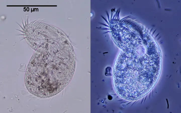
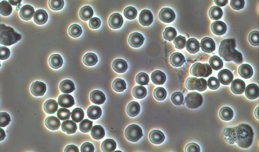
It is seen that a phase-contrast microscope is used for generating high contrast images of all types of biological samples. This technique is useful in a routine manner for the inspection of adherent cell cultures in most of the fields of biology and biomedicine. Despite the high contrasted images of all biological samples, it is a cumbersome process to adjust the contrast concerning the foreground and background. To eliminate this problem, the trainers and scientists use the process of segmentation in a pixel-wise manner. In the segmentation process of cells, the image structures and symmetries are encoded in the form of multi-scale basic images histograms. Here, further classification is done with the help of random decision trees proposed for the relevant phase microscopy images.
The segmentation process is validated for the cell versus background and discrimination between the two different types of cells. Phase contrast microscopy is a widely used microscopy technique that uses light properties. Microscopy modality of the inspection of adherent cell cultures is done by the phase-contrast microscopy techniques. The phase contrast microscopy is used in the cell magnification process in the role of de facto light. The observation of transparent cellular specimens is also done with the phase contrast microscopy technique. Here, the transformations of phase shifts are executed for magnifying the images of cultures.
In the proper principle of phase contrast microscopy, the different refractive indexes come into play that interfere with the source light and form the contrasted images. This leads to changes in the amplitude and refractive indexes of the induced light rays. All this can be detected by the human eye after the process of interference in the inverted phase contrast microscope. The type of segmentation is good for the formation of images in the inverted phase microscope for yielding high contrast images. Cell culture microscope, tissue culture microscope, and culture microscope using phase contrast microscopy techniques to magnify the biological images. A digital camera can also be fitted to the trinocular head of the microscope for recording the images for further observations.
Segmentation helps in the observational processes of biological images that are under the microscope. A challenge to automated segmentation of phase contrast microscopy images is the intrinsic structures of the artifacts of the biological images. However, this can be prevented by using histograms for the precision reading of images. In the process of segmentation, the shade-off effect results in the low contrast effects in the interior of cellular objects placed in the staging point of the trinocular compound microscope. The image background and bright halo artefacts can be seen beside the cellular objects in the cultures. Other sources of noise or disturbances are also seen in the process of segmentation of phase contrast microscopy illumination patterns with non-cellular backgrounds.
In this process, the phase contrast microscopy images are segmented based on the local histograms of BIFs (Binary Format For Scenes). These histograms are locally produced by the study of all neighbouring cells in the culture or an individual manner. Following are the steps used in the process of trainable segmentation in the PCM process :
The classification process depends on the formation of trees by the pixel methods which imparts segmentation to the biological samples. The number of trees had to be chosen by the researcher and scientists for taking into account the balance between the segmentation process and processing time. It is seen that the increasing number of trees above 20 showed only marginal improvement of segmentation performance. On the contrary, there is some increase in processing time with the increase in the number of trees in the phase-contrast images. With the processing time, memory usage also increases. On the other sides, the lower number of trees in the phase microscopy technique ensures reasonable processing times for the applications, where rapid feedback is required. In interactive segmentation, this process is done in the process of interactive segmentation.
The output of the classifier used in the segmentation processes is in the form of a binary label, 0 or 1 for foreground objects i.e, the biological cells. Although, the final judgment is based on the majority of trees formed by vectors used in the phase contrast microscopy. All these outputs are used for the segmentation without any further processing or refinement. A random Matlab software can be introduced in the segmentation process to create the results out of the biological samples.
BIFs computations are the backbone of the segmentation of the histograms used in phase contrast microscopy images techniques. The computations of BIFs consist of the classification of output obtained from the convolution of a biological image with a bank of derivatives of Gaussian filters. All these categories correspond to distinct local image structures which are defined by local symmetry slopes. These computations of BIFs give out symmetrical dark and bright lines with saddle points and flat colors. This makes the segmentation work easier for the respective histograms formed in the biological samples.
In addition to all BIFs features, the intensity and contrast features are also added to the segmentation process of all biological samples. For the intensity feature, the scales of the features correspond to the standard deviation of the Gaussian kernel is used in the segmentation process. The Gaussian kernel is used to blur the original phase contrast microscopy image. As per the process of intensity feature, the computations are fed into the respective computer for the phase contrast microscope. This results in a soft-edged normalized contrast filter.
This feature scale corresponds to the standard deviation of the concerned filter. Here, the local contrast histograms for both intensity and contrast features were constructed for giving out the high contrasted biological images.
Conclusion
The best trinocular microscope with a phase contrast feature is best for the segmentation process. Segmentation procedures are computed into the centered software for giving out the best phase contrast images. One can check the trinocular microscope price online as well as the offline medium for choosing the right microscope setup for the laboratory.