Sample Preparation
Homogenization
Heating and Mixing
Electrophoresis and Blotting
Polyacrylamide Gel Electrophoresis
Agarose Gel Electrophoresis
Western Blotting
Power Supplies
PCR & qPCR Thermal Cycler
Thermal Cycler (PCR)
Real-time Thermal Cycler (qPCR)
PCR Workstations & Cabinets
UVP BioImaging Systems
Molecular Spectroscopy
Lab Equipment
Ultraviolet Products
Hybridization Ovens
UVP Incubator
UV Crosslinkers
UVP Benchtop Transilluminators
Thermal Mixers
Electrophoresis & Blotting
Thermostats
View All
Fume hood
Laminar Airflow
Biosafety Cabinet
Autoclave
Centrifuge
pH Meter
Shaker & Mixer
Orbital Shaking Incubator
BOD Incubator
Heating Oven
Water Purification System
Aermax - Air Purification
Medical Oxygen Concetrators
Hygiene Solution
-150°C Cryogenic Freezer
-86°C Ultra Low Temp Freezer
-40°C Low Temp Freezer
-18 ~ -25°C Biomedical Freezer
-20°C Biomedical Freezer
4° ± 1°C Blood Bank Refrigerators
2~8°C Pharma Refrigerators
2~8°C ICE Lined Refrigerators
-25°C ~ + 4°C Mobile Freezer/Collers
20~24°C Blood Platelet Incubators
Ice Machines
Coldrooms
Mortuary Chambers
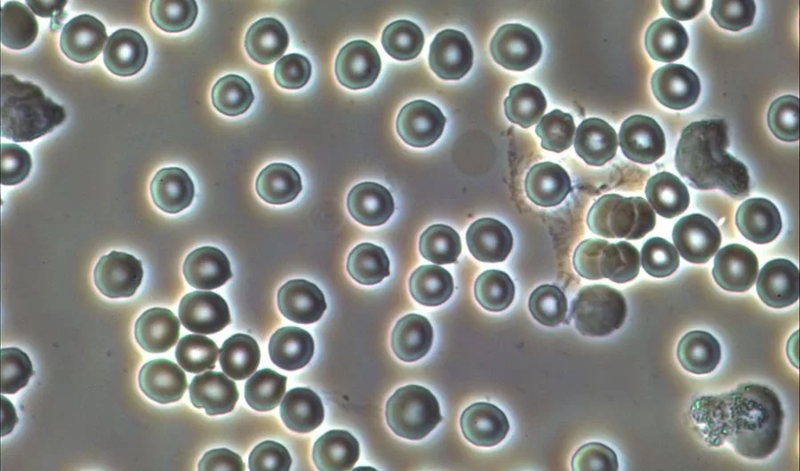
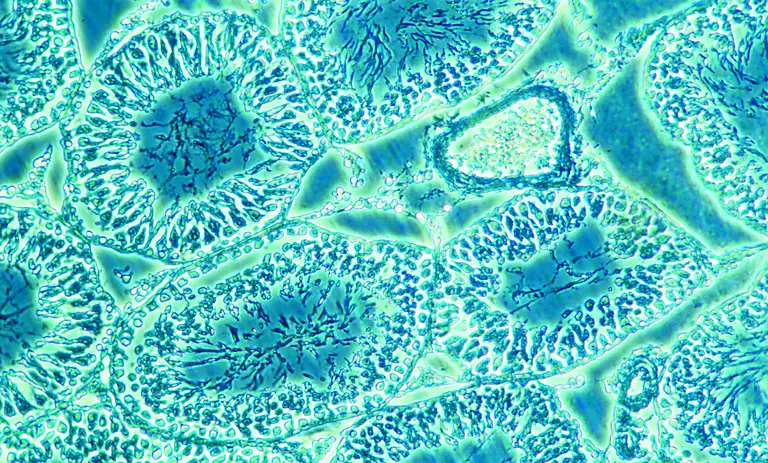
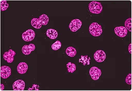
The microscopy technique has been in our scientific world since time immemorial. Many research labs and workplaces employ microscopes to get observations on cells, cultures, and many more microscopic creatures. Dark-field and phase contrast microscopes are the famous microscopy techniques for getting observational images of cells. Label-free imaging can happen with the dark field and phase-contrast microscopy. Here, the optical modes used are very accurate and subtle. With the help of the reflection and diffraction process of the electromagnetic property of light, the images can be obtained with full details. Dark-field microscopy techniques enable good contrast over the images obtained in the microscopy technique. An inverted microscope also can be coupled with the phase-contrast microscopy technique to become an inverted phase-contrast microscope.
The dark-field microscopy techniques generate images in their sub-resolution features that enable the perfect magnification. On the contrary, the phase microscopy techniques use the interferences of light rays to generate high contrast images. The high angle scattering of light rays happens in both types of microscopy techniques. Dark field microscopy is used for both unstained and stained objects, but the phase-contrast microscopy techniques are only targeted on the unstained phase objects. It is seen that many biological samples do not absorb the light rays, they are called phase objects in an inverted phase microscope. Both dark field and phase contrast microscopes can be used in the observations of cell culture, tissue culture, etc. Hence, they are called cell culture microscope, tissue culture microscope, and culture microscope. A wide range of light rays also gets scattered in both microscopy techniques which helps in generating detailed images. The upright and inverted microscope can also use dark field microscopy for good observations.
In scientific labs, the dark field microscope is used for the illumination of unstained biological samples. The technique helps the images to appear bright and fully contrasted images in the dark background. This dark background helps in the perfect illumination of all biological samples. All obtained images are full contrasted like images of phase contrast microscopy techniques. Dark field microscopes can be also used with the electron as well as light microscopy techniques. It has the feature of magnifying the small molecule to a larger resolution of pixels. A camera for a trinocular microscope can also be attached with the dark field microscope that can help in recording the images.
A dark field microscope has the following basic parts or components :
This condenser is called a special component of dark field microscopes. It scatters most of the light rays from the light source. The condenser also causes the light rays to reflect off the biological specimen at a certain angle. Here, the condenser does not illuminate the sample but fills the light rays to form a cone. The apex of the cone of light rays is all focussed on the plane of the biological specimen. This generates images of biological specimens.
The eyepiece is the small lens from where the researchers and scientists can observe the biological images of specimens used.
A light source is important for the magnification of biological specimens. The condenser used in the dark field microscope scatters the light rays if there is any light source. The whole scattering of light depends on the light source. In electron microscopy techniques, the light source is the high accelerating electrons only. The electrons help in the process of scattering light rays.
In a dark field microscope, the objective lens is situated at the dark hollows of the apex cone.
The light source showers the light on the microscope which gets scattered by the special condensers fitted into the microscope. As the light rays move past the biological specimen plane towards the apex of the hollow cone. The light pass on here moves to the hollow cones prepared by the reflected light. Also, the light rays travel past the objective lens in dark field microscopes, which seals the entry of other light rays in the trinocular compound microscope. The entire observational field appears dark when there is no biological specimen on the stage. But, when a biological specimen is placed on the stage, the light apex cone strikes it and generates the images. Hence, this microscopy technique is named a dark field microscope. All the rays that get scattered by the condensers are captured by the objective lens which further generates an image of the biological specimen.
In a phase-contrast device, it manipulates the light paths through the use of phase rings to illuminate transparent biological samples. Dutch physicist Fritz Zernike developed the phase-contrast technique in the 1930s and later he was awarded the Nobel prize in the same. In this technique, the parallel beams of light are passed through biological samples of different densities. The inverted phase microscope consists of special condensers that throw light “out of phase”. Here, the interferences in the wave nature of light occur. The rings cause it to pass through the object at different speeds. All Internal details of organelles of live, unstained organisms (e.g. mitochondria, lysosomes, Golgi body) can be seen clearly with a phase-contrast microscope. Even the best trinocular microscope also has the feature of interference to cause the generation of phase contrast images.
The basic components of a phase-contrast microscope are as follows :
The phase ring allows the light rays to pass through it while still in phase nature. Unaltered light rays hit the phase ring in the lens and are excluded later. The Light rays that are mildly altered by passing through a different refractive index are allowed to pass through the specimen. The Light rays passing through any type of cellular structure, such as chromosomes or mitochondria are retarded because they have a higher refractive index than the surrounding medium. Elements of a lower refractive index advance the waves of light rays. Most of the background light rays are removed. These light rays are let through with enhanced contrast. This whole process generates a high contrast image of biological specimens placed on the pointing stage.
The pointing stage is the place in the inverted phase-contrast microscope that holds the biological specimen.
The objective lens mediates the light rays to the plane of the biological sample for causing the interferences.
The phase-contrast microscopy technique allows the visualization of living cells in their natural state only. With high contrast and high resolution, all obtained images are detailed and good for observations. As the phase-contrast microscope gives out the best images, it comes a little more expensive than another inverted microscope. So, you can check the phase-contrast trinocular microscope price both online as well as offline. This microscope works best with a thin biological specimen and is not ideal for a thick specimen. All phase-contrast images have a characteristic grey background with light and dark expressions. This makes the whole process of observations a lot better and good.
Conclusion
Bioimaging is so important for any research lab. Here, the dark field microscope and phase-contrast microscope. Both works to give out high-resolution images of biological samples. The dark field microscope gives out the images through the light apex of the cone, while the phase-contrast microscope gives out highly contrasted images of the same. Both have their importance and are good for observational purposes. In research, bioimaging can be done with both of them.