Sample Preparation
Homogenization
Heating and Mixing
Electrophoresis and Blotting
Polyacrylamide Gel Electrophoresis
Agarose Gel Electrophoresis
Western Blotting
Power Supplies
PCR & qPCR Thermal Cycler
Thermal Cycler (PCR)
Real-time Thermal Cycler (qPCR)
PCR Workstations & Cabinets
UVP BioImaging Systems
Molecular Spectroscopy
Lab Equipment
Ultraviolet Products
Hybridization Ovens
UVP Incubator
UV Crosslinkers
UVP Benchtop Transilluminators
Thermal Mixers
Electrophoresis & Blotting
Thermostats
View All
Fume hood
Laminar Airflow
Biosafety Cabinet
Autoclave
Centrifuge
pH Meter
Shaker & Mixer
Orbital Shaking Incubator
BOD Incubator
Heating Oven
Water Purification System
Aermax - Air Purification
Medical Oxygen Concetrators
Hygiene Solution
-150°C Cryogenic Freezer
-86°C Ultra Low Temp Freezer
-40°C Low Temp Freezer
-18 ~ -25°C Biomedical Freezer
-20°C Biomedical Freezer
4° ± 1°C Blood Bank Refrigerators
2~8°C Pharma Refrigerators
2~8°C ICE Lined Refrigerators
-25°C ~ + 4°C Mobile Freezer/Collers
20~24°C Blood Platelet Incubators
Ice Machines
Coldrooms
Mortuary Chambers
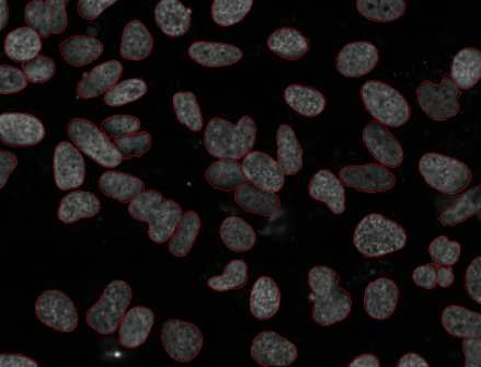
In the scientific world, there are many types of equipment that find use in the process of magnification and observation. Phase contrast microscopy, inverted microscope, culture microscope, all play a great role in the process of observation of bio-liquids and biomolecules. The origins of phase contrast microscopy are studied through the dynamic atomic force microscopy (dAFM) technique. In this technique, the low stiffness of microcantilever probes is manipulated which are famous for nanoscale bioimaging of soft biological specimens.
The forces used here are the gentle ones which are executed by the atomic force microscopy technique. It is observed that the phase contrast microscopy technique has its origin in atomic force microscopy. Under the above-mentioned conditions of gentle forces and probes, the phase contrast effect is derived from a primarily unique energy flow channel that opens up in bio-liquids due to its momentary excitation of higher eigenmodes. This is detected by the presence of contrasted images of samples that form under a trinocular compound microscope.
In other words, phase contrast images in liquids use soft microcantilevers which are often used for mapping of short-range conservative interactions in the biological specimens and liquids. These are the markings, such as the local elastic response of the sample tissues, rather than any tip-sample dissipation. This theory is used for the demonstration of variations in the local elasticity of purple membrane in the biomolecules and liquid. This technique is useful in the demonstration of bacteriophages in buffer solutions also by the phase contrast images.
Atomic force microscopy is the technique that generates contrasted images by scanning a small cantilever over the surface of a specimen. The trinocular head of the atomic force microscope uses the technique for producing images with high contrast. The process of microscopy leads the sharp tip on the end of the cantilever to contact the surface, which bends the cantilever and changes the amount of laser light reflected from the light source in an inverted phase microscope. Photodiodes are used as the light source for atomic force microscopy. When this microscopy technique is useful in the observations of cell cultures, they are called a cell culture microscope, or tissue culture microscope. Here, the height of the cantilever is adjusted to restore the response of the signal that results in the measured cantilever height tracing the surface of the biological specimens.
Atomic force microscopy (AFM) is also known as scanning force microscopy (SFM) that produces images of very high resolution. From here, the phase microscopy techniques got their origin to produce highly contrasted images. The small cantilever probes are scanned through the inverted phase contrast microscope, and then it is demonstrated for high resolutions in the order of fractions of a nanometer. The images produced by the scanning probe microscopy are more than 1000 times better than the optical diffraction limit of any traditional compound microscope. Mechanical probes are also used in the process of atomic force microscopy to form images. Piezoelectric elements are the elements that facilitate tiny but accurate precise movements in scanning microscopes. From the nature of atomic force microscopy, phase contrast microscopes originated. Today, for liquid imaging, phase contrast atomic force microscopes are used.
A normal atomic force microscope contains the following main components:
When the tip of the cantilever is brought into proximity surface, the forces between the tips come into play over the biological liquid sample. Here, the deflections of the cantilever happen according to Hooke’s law. In this situation, the forces measured in the atomic force microscopy include contact forces, capillary forces, van der waals forces, chemical forces, electrostatic forces, magnetic forces. etc. These all forces come into play and generate the images using contrast. Additional forces like solvation forces, casmier forces, also come into play for the formation of different contrasted images under the inverted phase-contrast images. Along with all these forces, some additional quantities also play their role in the image formation and scanning of all types of probes used in scanning thermal microscopy or expansion microscopy. The best trinocular microscope works by the atomic force microscopy technique to produce high contrasted and high-resolution images.
Dynamic Atomic Force Microscopy
Dynamic atomic force microscopy (dAFM) is an essential experimental tool for studying the conservation and dissipative force on the surfaces of biological samples. In this study, the results are obtained in the order of nanometers length scales. The results have major implications for physical biomolecular interactions within the samples. In addition to these, the adhesion, chemical bond kinetics, wetting, and capillary actions, and the elasticity of the biological specimens are taken into consideration. Dynamic atomic force microscopy has been developed by keeping all these biomolecular interactions in consideration which distinguishes between the dissipative forces (friction, bond-breaking, viscoelastic, surface hysteresis, capillary condensation) and the conservation forces (magnetic, electrostatic, elastic ). All these forces keep on oscillating between a sharp oscillating tip and the surface.
In the case of amplitude-modulated atomic force microscopy (AM-AFM) with phase contrast imaging, the phase variations keep on oscillating the probe tip concerning the drive signal that is mapped over the biological sample. In prior time, phase contrast microscopy techniques were connected closely with variations in tip-sample dissipation over the concerned sample. This modern world has recognized phase contrast microscopy techniques as the most important AM -AFM mode of measurement of compositional contrast.
The connection established between phase contrast and tip-sample dissipation stands on an assumption. As per the assumption, the cantilever dynamics can be modeled by a single eigenmode called a point-mass oscillator. Taking this assumption into consideration, the tip-sample dissipation can be equated to the difference between the work input to the oscillator and energy dissipated into the surrounding medium which is viscous. These facts or theories form the bedrock foundation upon phase-contrast imaging which is currently based on at least under ambient and vacuum conditions.
Dynamic atomic force microscopy is now a well-known microscopy technique that is a broadly extended technique for nanoscale imaging in spectroscopy methods. In the biology community, this process of magnification is very famous for its production of high contrasted images. This is because the natural medium for the study of all types of biological samples is liquid which is of fundamental importance to develop a proper description of different working modes of dynamic atomic force microscopy. In this mode, the probes and samples are immersed in liquids and then studied for phase differences.
It is seen that only a little is available for the understanding of the origins of phase contrast in liquids. Soft cantilevers of stiffness 1 N/m with low-quality factors are routinely employed for the imaging of soft biological samples. It is well understood that all types of theoretical works on phase contrast in air and liquids are based on a single eigenmode which is sufficient to describe the microcantilever dynamics.
Conclusion
The atomic force microscopy leads to the origins of the phase contrast microscopy techniques. In dynamic atomic microscopy techniques, the dissipations and the biomolecular interactions are taken into consideration to produce the high contrasted images of all types of biological samples.