Sample Preparation
Homogenization
Heating and Mixing
Electrophoresis and Blotting
Polyacrylamide Gel Electrophoresis
Agarose Gel Electrophoresis
Western Blotting
Power Supplies
PCR & qPCR Thermal Cycler
Thermal Cycler (PCR)
Real-time Thermal Cycler (qPCR)
PCR Workstations & Cabinets
UVP BioImaging Systems
Molecular Spectroscopy
Lab Equipment
Ultraviolet Products
Hybridization Ovens
UVP Incubator
UV Crosslinkers
UVP Benchtop Transilluminators
Thermal Mixers
Electrophoresis & Blotting
Thermostats
View All
Fume hood
Laminar Airflow
Biosafety Cabinet
Autoclave
Centrifuge
pH Meter
Shaker & Mixer
Orbital Shaking Incubator
BOD Incubator
Heating Oven
Water Purification System
Aermax - Air Purification
Medical Oxygen Concetrators
Hygiene Solution
-150°C Cryogenic Freezer
-86°C Ultra Low Temp Freezer
-40°C Low Temp Freezer
-18 ~ -25°C Biomedical Freezer
-20°C Biomedical Freezer
4° ± 1°C Blood Bank Refrigerators
2~8°C Pharma Refrigerators
2~8°C ICE Lined Refrigerators
-25°C ~ + 4°C Mobile Freezer/Collers
20~24°C Blood Platelet Incubators
Ice Machines
Coldrooms
Mortuary Chambers
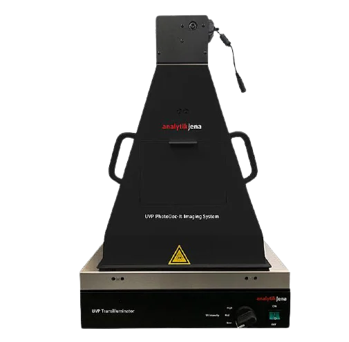
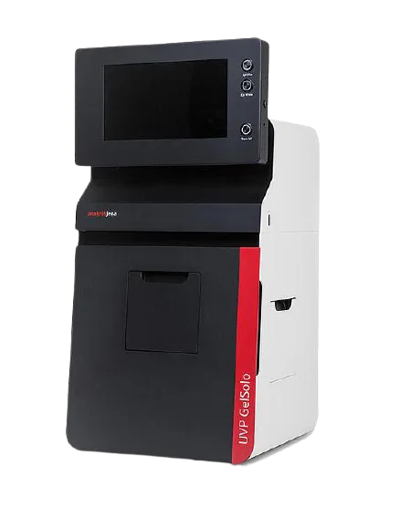
The Gel documentation system is an imaging system used to analyze proteins, nucleic acid, and antibodies suspended within polyacrylamide or agarose gels. Gel documentation systems or gel imaging systems are widely used in molecular biology laboratories.
Gel documentation works in the principle of fluorescence. Nuclei acids stained with a fluorescent substance such as Ethidium Bromide are excited by ultraviolet irradiation and emit fluorescent light.
Depending on the molecular weight and concentration of nucleic acid the amount of Ethidium Bromide binding is made specifically to nucleic acid.
In other words, the larger the molecular weight of nucleic acid, the higher the binding, and the fluorescence will shine brighter. Lower the molecular weight of nucleic acid, fluorescence will be weaker.
Read more : Gel Documentation System Procedure
Gel doc systems are commonly used in molecular biology, microbiology labs, DNA quantification, Gel imaging systems support PCR segment identification, bacterial cell culture, environmental sample testing, and protein separation.
Nucleic acid (DNA or RNA) samples are suspended within the acrylamide or agarose gel and when connected to the electric field, the negatively charged samples are attracted towards the positive electrode.
Smaller nucleic acid fragments travel faster than longer nucleic acid fragments, allowing researchers to identify the fragments with fluorescent dyes. DNA gels are generally used for next-generation sequencing, PCR analysis, and cloning.
Protein samples are suspended within polyacrylamide that is submerged in a buffer such as Tri-Glycine or Tri-Acetate. Protein fragments of different lengths will travel at different speeds through a gel matrix similar to DNA gels. These protein gels are commonly are used for mass spectrometry, blotting, and sample denaturing.
The process of determining the number of Colony Forming Units (CFUs) that are present in the petri dish suspended in the cell media under optimal growth conditions. Colony counting is used to determine microbes in the soil, water, or food samples and also to identify unique cell lines for microbial cell culture research.
Thin Layer Chromatography (TLC) is a research method through which the components of a mixture using a thin film is separated by an inert backing.
As the mixture moves through the stationary phases that include silica gel, cellulose, and aluminum oxide, the components move through the solvent (mobile phase) that includes methanol, acetic acid, and ethyl acetate.
Thin-layer chromatography is widely used to quantify and qualify many substances including lipids, carbohydrates, fatty acids, and pesticides.
Western blotting gels are used to separate proteins for isolation and purification. These western blotting gels are in two primary forms: native gels and SDS-PAGE gels.
For the study of enzyme or protein complexes, and to separate proteins based on size and ionic charge, native gels are used. SDS-PAGE gels are used to separate proteins based on size by denaturing samples using detergent such as Sodium Dodecyl Sulphate (SDS).
Southern blotting gels are used for the separation of DNA fragments from blood samples from the restrictive digestive enzymes. These gels are available in two forms polyacrylamide gels with urea and Sodium Dodecyl Sulfate (SDS) gels with urea.
Gel documentation or gel imager includes CCD or CMOS camera. A camera with a higher resolution (pixel count) will provide greater sensitivity and better image clarity. Generally, Gel documentation image comes with a resolution from 5 megapixels to 20 megapixels.
Different types of gel doc systems or gel imaging systems are used depending on the laboratory applications and detection methods.
Chemiluminescence is basically used for western blotting.
Flim Auto Radiography is used for radioisotopes
Laser is used for multiplex fluorescence applications, high sensitivity, and precise quantitation for radioisotopes
Multiplex is used to detect and image multiple fluorescent signals simultaneously. This type of gel imaging system is also used to detect luminescent, colorimetric, and radioisotopic signals.
Phosphoimagers is the advance of multiplex fluorescence applications and radioisotopes It is more sensitive than multiplex gel imagers. It is more flexible in choosing emission filters.
CCD/ Digital Ethidium Bromide (UV), infrared, color fluorescence, densometric, chemiluminescence, and visible light detectors are available to take quantitative reads of nucleic acid, protein fragments, microplates, and dot blots.
Other Applications of Gel Doc System:
Conclusion
A gel documentation system is a basic requirement for all labs that relates to molecular biology. The gel documentation system or gel imager is available commercially in different types depending on different types of applications. It is important to choose the right gel documentation for the lab applications.