Sample Preparation
Homogenization
Heating and Mixing
Electrophoresis and Blotting
Polyacrylamide Gel Electrophoresis
Agarose Gel Electrophoresis
Western Blotting
Power Supplies
PCR & qPCR Thermal Cycler
Thermal Cycler (PCR)
Real-time Thermal Cycler (qPCR)
PCR Workstations & Cabinets
UVP BioImaging Systems
Molecular Spectroscopy
Lab Equipment
Ultraviolet Products
Hybridization Ovens
UVP Incubator
UV Crosslinkers
UVP Benchtop Transilluminators
Thermal Mixers
Electrophoresis & Blotting
Thermostats
View All
Fume hood
Laminar Airflow
Biosafety Cabinet
Autoclave
Centrifuge
pH Meter
Shaker & Mixer
Orbital Shaking Incubator
BOD Incubator
Heating Oven
Water Purification System
Aermax - Air Purification
Medical Oxygen Concetrators
Hygiene Solution
-150°C Cryogenic Freezer
-86°C Ultra Low Temp Freezer
-40°C Low Temp Freezer
-18 ~ -25°C Biomedical Freezer
-20°C Biomedical Freezer
4° ± 1°C Blood Bank Refrigerators
2~8°C Pharma Refrigerators
2~8°C ICE Lined Refrigerators
-25°C ~ + 4°C Mobile Freezer/Collers
20~24°C Blood Platelet Incubators
Ice Machines
Coldrooms
Mortuary Chambers
During fluorescent staining of nucleic acids, a fluorescent substance (ethidium bromide) is bound to nucleic acids, is excited by ultraviolet irradiation, and emits fluorescent light. The fluorescent substance binds specifically to nucleic acid and the amount of binding is dependent on the concentration and molecular weight of the nucleic acid. So, a band for a large molecular weight or a large amount of sample will shine more luminous; contrarily, fluorescence will be weaker for a band in the case of smaller molecular weight or a tiny quantity of analyte. With continued irradiation of UV rays (254 nm, a short wavelength), the fluorescence of a band weakens gradually. This is imminent when the molecular weight or the amount of the sample is small.
Ethidium bromide binds to double-stranded DNA by inserting between grooves of stacked bases. The ring structure of ethidium is hydrophobic and it also resembles the rings of the bases present in DNA. Ethidium bromide is able to form close van der waals interactions with the base pairs and hence, it binds to the hydrophobic interior of the DNA molecule. It intercalates in the stacked base pairs of DNA and elongates DNA by untwisting the helix. Ethidium bromide is a fluorescent molecule. It is a well-known fluorescent tag. When illuminated with visible light, it is clearly visible. This makes contents to be visualized using a transilluminator. This principle is used in techniques like agarose gel electrophoresis for viewing proteins and nucleic acids. Ethidium bromide is abbreviated as EtBr and sometimes as EthBr.
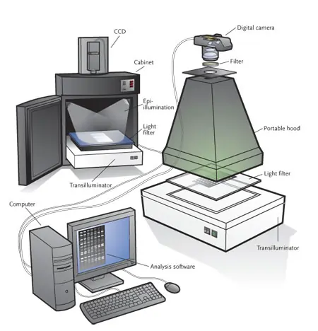
Ethidium bromide is useful in detecting nucleic acids in genetic engineering and molecular biology laboratories using the gel doc system. In the case of DNA, this is usually double-stranded DNA from PCRs, restriction digests. Single-stranded RNA can also be detected, as it usually folds back onto itself, and thus local base pairing for the dye to intercalate takes place. In detection, the gel containing nucleic acids is placed under a UV light. As UV light is harmful to skin and eyes and ethidium bromide can be a mutagen depending on the exposure time, gels stained with ethidium bromide are generally viewed indirectly with an enclosed camera and the fluorescent images are recorded as photographs in a computer. If direct viewing is required, the eyes and exposed skin of viewers should be protected. Generally, stock solutions of ethidium bromide solutions are prepared at 0.25-1.0 microgram/mL concentration for the purpose of gel staining. Ethidium bromide is useful during the separation of DNA fragments by agarose gel electrophoresis. It is added to the running buffer and it binds by intercalating between DNA base pairs. When the gel is illuminated using UV light, DNA bands become visible.
Ethidium bromide helps in the separation of closed circular DNA molecules and linear DNA (genomic DNA). It cannot bind to closed molecules because it is not able to get inserted between the bases as the length is lesser. Also, it increases the density of DNA upon binding, thus paving way for easy separation between circular and linear DNA.
One molecule of ethidium bromide is intercalated per 2.5 base pairs of DNA. Absorption peaks of EtBr (360 nm and 300 nm) fall inside the UV range of the spectrum. Ethidium bromide emits orange light with a wavelength of 605 nm upon excitation. Besides, it also absorbs energy from nucleotides, which are excited by the absorbance of 260 nm radiation. Ethidium re-emits this energy as yellow/orange light at 590 nm. In an aqueous solution, the fluorescence exhibited by ethidium bromide is significantly lesser than that of the intercalated dye.
Ethidium bromide is a cationic dye. In addition to fluorescence enhancement, there are some other sites involved which are fluorescence quenching and exhibit electrostatic binding. The equilibrium conformation and solvent environment of the dye have been modified on interaction with DNA. The ethyl and phenyl groups present in the peripheral region inserted into the major groove of the DNA helix. When the gel is bound with ethidium bromide, the mobility of DNA is mitigated.
The change in frictional coefficients by the addition of ethidium bromide is directly proportional to the number of base pairs of a DNA fragment bound to ethidium bromide. In longer fragments, the change in friction is greater, which implies that the stiffening of the DNA molecule by binding of ethidium bromide is the cause for the decreased mobility. Intercalation of EtBr is capable of changing the properties of DNA, like weight, charge, stability, conformation, and flexibility. As the mobilities of DNA molecules through the agarose gel are usually measured in relation to a molecular weight standard, the impact of EtBr can be significant to determine the sizes of DNA molecules.
Conclusion
The fluorescence phenomenon is behind the gel documentation system. A fluorescent dye is used as a marker for the visualization of DNA or protein molecules loaded into the gel. For this process, ethidium bromide dye is indispensable as it is used in most laboratories. If any concern arises in the safety of ethidium bromide and to save the time required for decontamination, it is recommended to use other fluorescent gel SYBR Safe, Crystal Violet.