Sample Preparation
Homogenization
Heating and Mixing
Electrophoresis and Blotting
Polyacrylamide Gel Electrophoresis
Agarose Gel Electrophoresis
Western Blotting
Power Supplies
PCR & qPCR Thermal Cycler
Thermal Cycler (PCR)
Real-time Thermal Cycler (qPCR)
PCR Workstations & Cabinets
UVP BioImaging Systems
Molecular Spectroscopy
Lab Equipment
Ultraviolet Products
Hybridization Ovens
UVP Incubator
UV Crosslinkers
UVP Benchtop Transilluminators
Thermal Mixers
Electrophoresis & Blotting
Thermostats
View All
Fume hood
Laminar Airflow
Biosafety Cabinet
Autoclave
Centrifuge
pH Meter
Shaker & Mixer
Orbital Shaking Incubator
BOD Incubator
Heating Oven
Water Purification System
Aermax - Air Purification
Medical Oxygen Concetrators
Hygiene Solution
-150°C Cryogenic Freezer
-86°C Ultra Low Temp Freezer
-40°C Low Temp Freezer
-18 ~ -25°C Biomedical Freezer
-20°C Biomedical Freezer
4° ± 1°C Blood Bank Refrigerators
2~8°C Pharma Refrigerators
2~8°C ICE Lined Refrigerators
-25°C ~ + 4°C Mobile Freezer/Collers
20~24°C Blood Platelet Incubators
Ice Machines
Coldrooms
Mortuary Chambers
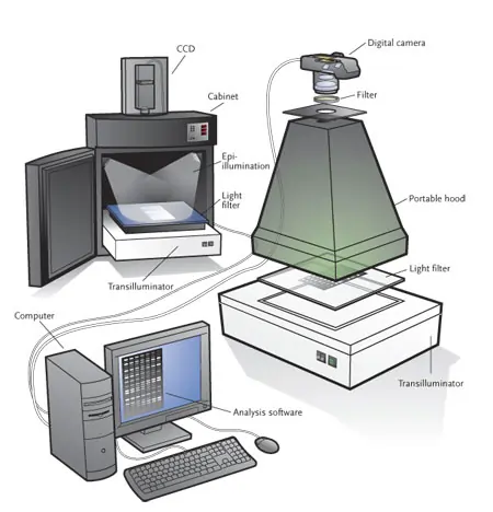
A gel documentation system is a system used primarily in molecular biology labs with the objective of imaging and documentation nucleic acids and proteins, suspended in agarose or polyacrylamide gels stained with fluorophores like SYBR green or ethidium bromide. A gel doc system contains a light source, transilluminator, CCTV camera for capturing the image, and a dark room or hood to shield the body from UV light. It is also known as gel imager, gel image system, and gel doc system.
Gel documentation systems are used in genetic engineering laboratories in colleges, genetic engineering research institutes and protein-producing pharmaceutical industries. It is also used in forensic laboratories and clinical research labs.
Gel imaging or gel documentation System is a system for recording and measuring labeled protein and nucleic acid in different media like acrylamide, cellulose, or agarose. It is used in the rapid evaluation of gels post electrophoresis, thus saving chemicals and time. It leads to visualization in less than 5 minutes after electrophoresis. Besides, it employs stain-free technology.
Depending on the type of sample type and throughput, Different configurations and specifications come in the gel doc system.
The different systems are as follows:
Chemiluminescence Imaging System
In the life science laboratory, western blotting is an important technique for the separation of proteins based on molecular weight. Chemiluminescence is used as a detection method for a western blot on account of high sensitivity, and imaging systems with this technique further optimize speed, sensitivity, and signal stability.
Digital Gel Imaging Systems
Digital gel imaging systems are being used to record and measure stained acrylamide and agarose gels on a digital platform. They not only provide effective data storage, but also accurate quantification of samples. Different types of detectors (visible light, Ethidium bromide (UV), fluorescence, chemiluminescence and, densitometric detectors) are available for quantitative analysis of protein and nucleic acid bands, microplates and dot blots. This system simplifies the image acquisition process with options of auto-exposure, and auto-focusing.
Automated Blot Analysis
Automated blot analysis systems are sensitive imagers, which utilize specialized software in analyzing western and nucleic acid blot data. These systems are capable of doing automation like focusing, light selection, exposure, and acquisition. Choices in detection range from UV, infrared, RGB color, fluorescence, chemiluminescence. They have multiple fluorescent channels, which lead to the measurement of multiple targets simultaneously.
Multiplex Fluorescence Imaging Systems
Multiplex fluorescent imaging systems are employed in illumination of blotting membranes, and detection of the signals over background noise. They are also involved in capturing the blot image and analysis of the signals. Most systems contain a light source, emission filters, and a photosensor (CCD or CMOS camera). Probable light sources are RGB, UV, and white light. Some systems are able to execute multiplexing capabilities to detect and image multiple fluorescent signals simultaneously. Some fluorescent imaging systems can detect radioisotopic, luminescent, and colorimetric signals. Some systems will use the principle of confocal optics to boost sensitivity when detecting targets present in low concentration.
Gels used in Electrophoresis
Gel electrophoresis is a technique in the laboratory that enables the separation and analysis of charged molecules in an electric field. The Separation is based on the size or conformation of molecules in a matrix, which is made from gel-forming components. Polyacrylamide gel electrophoresis and agarose gel electrophoresis are typically used as they are compatible with different sizes and types of the analyte. Agarose is apt to separate fragments of DNA, which range in size from a few 100 base pairs to around 20 kb. Polyacrylamide gel is preferred for smaller DNA fragments and proteins. Initially, starch gel prepared from soluble potatoes was used for the electrophoresis. But, due to the occurrence of batch-to-batch variations, agarose and polyacrylamide gels have replaced starch.
There are some modifications to gel electrophoresis nowadays.
There are numerous advantages in the use of the gel doc system.
There are numerous applications of the gel doc System, namely
There are some factors, which are needed to analyse before purchasing a gel doc system. They are as follows:
Conclusion
A gel documentation system is a prerequisite for any lab involving protein analysis. It comes in a diverse configuration and specifications with an option of customization depending on the level of application. As funding and budget are essential for any laboratory, it is necessary to purchase the best one tailored to each laboratory's needs. Utmost care should be taken to know about the after-sales service quality of the company for the equipment.