Sample Preparation
Homogenization
Heating and Mixing
Electrophoresis and Blotting
Polyacrylamide Gel Electrophoresis
Agarose Gel Electrophoresis
Western Blotting
Power Supplies
PCR & qPCR Thermal Cycler
Thermal Cycler (PCR)
Real-time Thermal Cycler (qPCR)
PCR Workstations & Cabinets
UVP BioImaging Systems
Molecular Spectroscopy
Lab Equipment
Ultraviolet Products
Hybridization Ovens
UVP Incubator
UV Crosslinkers
UVP Benchtop Transilluminators
Thermal Mixers
Electrophoresis & Blotting
Thermostats
View All
Fume hood
Laminar Airflow
Biosafety Cabinet
Autoclave
Centrifuge
pH Meter
Shaker & Mixer
Orbital Shaking Incubator
BOD Incubator
Heating Oven
Water Purification System
Aermax - Air Purification
Medical Oxygen Concetrators
Hygiene Solution
-150°C Cryogenic Freezer
-86°C Ultra Low Temp Freezer
-40°C Low Temp Freezer
-18 ~ -25°C Biomedical Freezer
-20°C Biomedical Freezer
4° ± 1°C Blood Bank Refrigerators
2~8°C Pharma Refrigerators
2~8°C ICE Lined Refrigerators
-25°C ~ + 4°C Mobile Freezer/Collers
20~24°C Blood Platelet Incubators
Ice Machines
Coldrooms
Mortuary Chambers
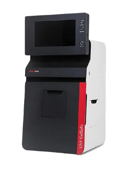
A gel documentation system is a system used primarily in molecular biology labs with the objective of imaging and documentation nucleic acids and proteins, suspended in agarose or polyacrylamide gels stained with fluorophores like SYBR green or ethidium bromide. It is also known as gel imager, gel imaging system, and gel doc system.
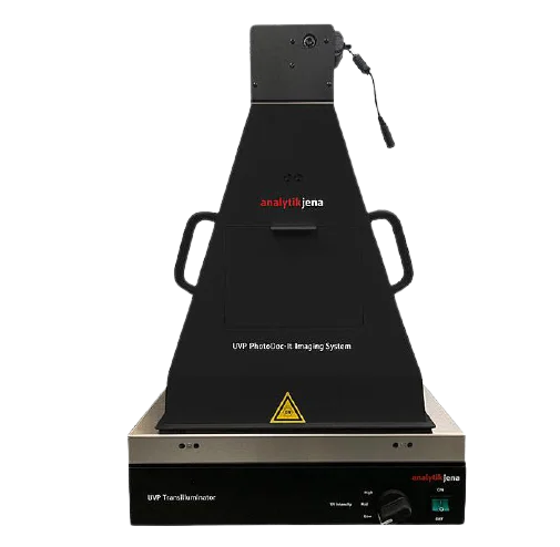
Transilluminator is the source of irradiation and is used in the observation of DNA bands. Usually, UV light is used for radiating samples. It is used when ethidium bromide is used as a fluorescent tag in samples obtained from agarose gel electrophoresis. UV lights may be four in number for use. The excitation range can vary from 250-800 nm. Shut off timer can be activated after a certain period of inactivity of the source. The latest technology involves no need to warm up UV lamps and there is instant switching on.
Nowadays, white light and LED lighting options are also available. It has the option of variable intensity setting. There may be safety cut-off values to prevent damage to the sample and to the operator. The fluorophore or fluorescent tags used in the gel should correspond to the irradiation source. This selection helps in choosing high performance or cost-cutting. If there is lee funding, UV to visible converter plate can be used instead of purchasing visible light source.
The base plate is a sample tray and can be easily pulled out. It is used to hold the gel for viewing. It is placed on a non-reflective and black surface.
It is a darkroom for shielding the skin against UV rays. It is suitable for fluorescence, chemiluminescence, and visible light applications. There is an electronic auto door lock, which is used for the prevention of longer exposures to radiation. It ensures proper documentation of captured images by acting as a dark box. Fold down door and slide-to-door options are available for less interference.
Amber filters should be able to shield the UV radiation fully from the eyes. It is also used in blocking background light, otherwise, the noise will appear on captured images. Emission filters are used to block the UV radiation for safe viewing of the illumination of the sample. An extensive variety of filters are available depending on the type of application.
Visible light is used for the extension of transmitted light application. For visible light application, visible light converts the screen.
It is used to move the sample closer to the camera.
It has a higher resolution camera. The camera controller and printer are also there. The resolution of the camera normally ranges from 1.4 MP to 8.3 MP. Flatbed scanners and CCD (Charge coupled device) cameras are used. Lens are motor driven and computer controlled. Using the option of auto-focus, lenses can track the downward and upward movements of samples automatically. Before capturing images, it is adjusted as it is directly connected with the readout system.
The camera is present above the hood for capturing images. Effective photon to signal conversion and deep cooling technologies are available. In low-light applications, a wide aperture lens is used to capture more light. The camera will satisfy a wider dynamic range and maximum light sensitivity.
The computer system has a conventional monitor or touch screen. Software should be available for computer-controlled capture and editing of images, This part is used in optimizing, quantifying, and storing the data obtained in a precise and accurate manner. This can be done by an integrated monitor of the system or a separate laptop connected to the system. Software installed is used for the optimization of image acquisition and analysis. Image enhancement tools are used for background subtraction, correction of the dark frame, noise removal, and inversion. USB ports are available for copying and transferring data to other systems.
Macros and templates are useful to create workflows with a single touch. It is used for reading and documenting colorimetry, gels, western blots, plants, TLC plates, colony blots, fluorescent dyes, and infrared dyes. A variety of tools are available for the analysis of data. Simultaneous imaging of multiple blots and gels is available for the gel to gel comparison. For calculation, the calibration standard is made precise and there is an accurate determination of concentration. A solid-state drive or hard disk drive is available for the storage of large volumes of data, templates, and images. Besides, through wi-fi and wired-to-ethernet connection, the system can be used for massive sharing of data immediately. The operating system can be a user-friendly version of windows.
Conclusion
From the above points, we come to know about the different components present in a gel documentation system. Each part is indispensable and has a function, contributing to the imaging and documentation of gels obtained from electrophoresis. The gel imager finds its application in post-translational modifications, blotting technique analysis, and 2-D electrophoresis documentation also.