Sample Preparation
Homogenization
Heating and Mixing
Electrophoresis and Blotting
Polyacrylamide Gel Electrophoresis
Agarose Gel Electrophoresis
Western Blotting
Power Supplies
PCR & qPCR Thermal Cycler
Thermal Cycler (PCR)
Real-time Thermal Cycler (qPCR)
PCR Workstations & Cabinets
UVP BioImaging Systems
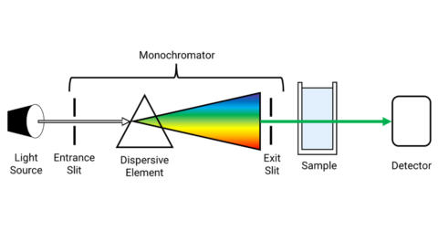
Molecular Spectroscopy
Lab Equipment
Ultraviolet Products
Hybridization Ovens
UVP Incubator
UV Crosslinkers
UVP Benchtop Transilluminators
Thermal Mixers
Electrophoresis & Blotting
Thermostats
View All
Fume hood
Laminar Airflow
Biosafety Cabinet
Autoclave
Centrifuge
pH Meter
Shaker & Mixer
Orbital Shaking Incubator
BOD Incubator
Heating Oven
Water Purification System
Aermax - Air Purification
Medical Oxygen Concetrators
Hygiene Solution
-150°C Cryogenic Freezer
-86°C Ultra Low Temp Freezer
-40°C Low Temp Freezer
-18 ~ -25°C Biomedical Freezer
-20°C Biomedical Freezer
4° ± 1°C Blood Bank Refrigerators
2~8°C Pharma Refrigerators
2~8°C ICE Lined Refrigerators
-25°C ~ + 4°C Mobile Freezer/Collers
20~24°C Blood Platelet Incubators
Ice Machines
Coldrooms
Mortuary Chambers
Raman spectroscopy is a laboratory analytical technique in which scattered light is used to measure the vibrational energy modes of a sample. This technique is named after the renowned Indian physicist C. V. Raman, who was the first person to observe Raman scattering or Raman shift in 1928 along with his research partner K. S. Krishnan.
This article takes you to the different variants of Raman imaging. Some of them are enhanced techniques. Their working principle and usage have been discussed.
More than 25 various types of Raman spectroscopy imaging techniques have been discovered. Among them, some have emerged popular. Let us see some major techniques.
Surface plasmon polaritons (SPP) are electromagnetic modes, which are propagating at the interface between a negative and positive permittivity medium because of resonant oscillations of free carriers. They have the characteristic to confine photon energy into subwavelength volumes at the conductive or dielectric interface where the electromagnetic evanescent field is strongly increased. This property has increased the detection and imaging capabilities. So, SPPs are utilized to perform remotely excited SERS, which results in a technique where excitation and SERS signal collection are displaced spatially. This reduces the origination of fluorescence background signals. Thus, the term Surface Plasmon Polariton Enhanced Raman Scattering (SPPERS) has been derived.
SERRS is a selective and sensitive method used for the characterization of sites in biomolecules, which possess an electronic transition at an energy close to the laser frequency used. Here, the sensitivity of resonance with that of the SERS technique is combined, so very low concentrations can be used.
The cons are:
Resonance Raman Spectroscopy
The table below shows the pros and cons of some variants of Raman spectroscopy:
Conclusion
We have come to know about different variants of Raman spectroscopy. Though all variants are not discussed, important types of Raman spectroscopy have been described. Some variants have become indispensable to advanced experiments. However, depending on the severity of the need, the variant is chosen and the Raman imaging process is executed.