Sample Preparation
Homogenization
Heating and Mixing
Electrophoresis and Blotting
Polyacrylamide Gel Electrophoresis
Agarose Gel Electrophoresis
Western Blotting
Power Supplies
PCR & qPCR Thermal Cycler
Thermal Cycler (PCR)
Real-time Thermal Cycler (qPCR)
PCR Workstations & Cabinets
UVP BioImaging Systems
Molecular Spectroscopy
Lab Equipment
Ultraviolet Products
Hybridization Ovens
UVP Incubator
UV Crosslinkers
UVP Benchtop Transilluminators
Thermal Mixers
Electrophoresis & Blotting
Thermostats
View All
Fume hood
Laminar Airflow
Biosafety Cabinet
Autoclave
Centrifuge
pH Meter
Shaker & Mixer
Orbital Shaking Incubator
BOD Incubator
Heating Oven
Water Purification System
Aermax - Air Purification
Medical Oxygen Concetrators
Hygiene Solution
-150°C Cryogenic Freezer
-86°C Ultra Low Temp Freezer
-40°C Low Temp Freezer
-18 ~ -25°C Biomedical Freezer
-20°C Biomedical Freezer
4° ± 1°C Blood Bank Refrigerators
2~8°C Pharma Refrigerators
2~8°C ICE Lined Refrigerators
-25°C ~ + 4°C Mobile Freezer/Collers
20~24°C Blood Platelet Incubators
Ice Machines
Coldrooms
Mortuary Chambers
A biology lab is full of experiments and observations which are carried daily. The study ranges from transparent objects to thick cultures. All types of observations are done using microscopes which are structured in a detailed way to observe the biological specimen. For many types of experiments, different microscopes are used which gives a detailed study of the specimen. When it comes to microscopes, people always imagine the conventional compound one which has a trinocular head and a pointing stage for the specimen. With the technical advancement in the microscopic apparatus, many biological microscopes were invented with full details to ace up the study of biological specimens. These microscopes are the extended version of the conventional trinocular compound microscope.
These microscopic devices are employed by researchers and scientists, and medical technicians daily to study and get insights into the biological specimens mounted on a microscope stage. Researchers tend to select the type of microscopes they need for their experiments. Some of the microscopes provide greater resolution, like phase contrast microscope, and some give out images of small resolutions, like simple upright and inverted microscopes. It is always advised to use the best trinocular microscope, for observing the cells and cultures.
As mentioned above, many types of microscopes are employed for deducing observational data from biological cells and tissues.
Following types of microscopes are used in a biology laboratory that help study the biological specimens :
Simple microscopes are generally considered the first microscope to be used for the observations. The microscope was created in the 17th century by combining a convex lens with a holder for the specimens used. This simple microscope’s magnifying power is between 200 and 300 times, which is good for the magnification of the biological specimens. The red blood cells were the first microscopic cells which are studied with a simple microscope. Today, simple microscopes are not that much used because of their low magnification power. However, the simple microscope is the base of all advanced microscopy techniques which we use in our daily biology experiments. This has led to the discovery of many powerful compound microscopes in the scientific world. Some transparent tissues or cells are well magnified in the simple microscope.
Compound microscopes are made up of two lenses which offer better magnification than the traditional trinocular microscope. Here, the second lens magnifies the images of the first lens. These compound microscopes are brightfield microscopes in which the specimen is lit underneath. The images formed in the trinocular compound microscope can be binocular or monocular.
The compound microscopes provide a magnification of 1,000 times more than the simple microscopes. But the resolution of the image formed is low, because of this, the compound microscopes are not that much in use. However, the compound microscope allows the users to take a closer look at the objects which are too small to be seen by the naked eye. Individual cells or aggregate of cells can also be magnified by the use of a compound microscope. The specimens used in the compound microscope’s magnification are relatively transparent than the other biological specimens. In a manner of cost-effectiveness, the compound microscope is a little more expensive than the simple microscopes. Because of its high power of magnification, it is used in biology labs for studying cultures and individual cells.
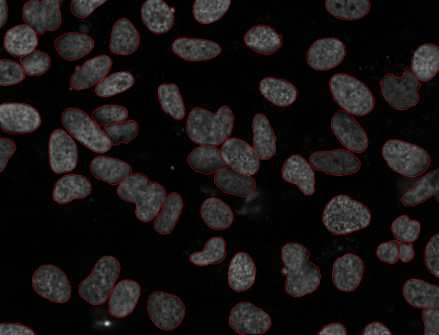
Unlike compound microscopes, confocal microscopes use regular light for the generation of images. In confocal microscopes, the light source used is a laser light that scans the biological samples that are labeled with the dye. These biological samples are labeled with the dyes, on the slides and mounted over the stage with the help of a dichromatic mirror. This mirror device helps with the generation of images of 3-D types by assembling the multiple scans in one place. The confocal microscopes show a high degree of image magnification, with good resolutions. All images formed are of good resolutions which are far better than simple and compound microscopes. Confocal microscopes are used mainly in cell biology and medical applications. These are also used to study tissue culture samples, so they are also called tissue culture microscopes and cell culture microscopes.
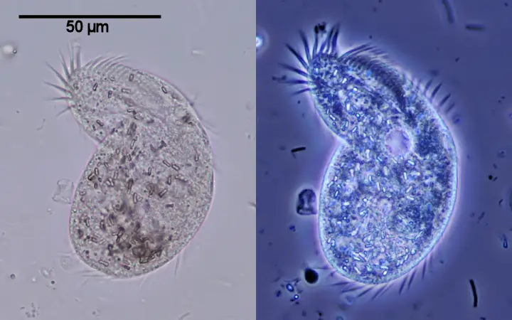
The scanning electron microscope or SEM is the new advancements in the field of microscopic image formation. In this technique, the samples are scanned in vacuum or near-vacuum conditions. This is done to make the samples well prepared for scanning purposes. The preparation is done by dehydrating the samples and then coating them with a thin layer of conductive material, like gold or any other metal. After this the item is placed on the pointing stage of the scanning electron microscope‘s chamber. The SEM produces a 3-D black and white image on the connected computer screen which gives a good insight into the sample. The samples can be seen through the computer monitor and can be used to examine the physical, medical, and biological phases of the insects and bones.
Transmission electron microscopes are somewhat the same as transmission microscopes. Here, also the electron beams are used for magnifying the biological images. The electron beams are used to create the magnified images of all types of biological specimens. All samples are scanned in a vacuum and specially prepared for the transmission electron microscope. A culture microscope also uses the principle of the transmission electron microscope (TEM) to produce images of great interest. The transmission electron microscope uses slide preparation to obtain a 2-D view of specimens which is well suited for the preparation of biological specimen images. The objects with the least transparency are used for the samples in the transmission electron microscope. The inverted microscope can also use the principle of TEM for generating the detailed structures of the specimens. A TEM-led microscopic device offers a high degree of magnification and resolution which is useful in the physical and biological sciences, nanotechnology, and metallurgy analysis.
Phase contrast microscopes came from the discovery of Frits who used the confocal lens to generate an illuminated image of the biological specimen. In phase contrast microscopy, the phase differences of light are used very deliberately to give out the illuminated image. The different refractive indexes of the surface of the biological specimen pave the way for the phase differences which help in generating images of the biological samples. Here, the wave nature of light is fully exploited to bring out the images of the specimen. All images formed are of illuminating nature that helps the researchers and scientists to have a close look-up over the surface of cells and cultures.
Conclusion
The best trinocular microscope has any of the above microscopy techniques which helps in image formation. Simple and compound microscopes are the primitive microscopy techniques that have become the base of many advancements in microscopy techniques. Transmission and scanning electron microscopy techniques are way too advanced in generating high-resolution and magnified images of all types of biological specimens. The inverted microscope price can be checked online or offline at the user’s convenience.