Sample Preparation
Homogenization
Heating and Mixing
Electrophoresis and Blotting
Polyacrylamide Gel Electrophoresis
Agarose Gel Electrophoresis
Western Blotting
Power Supplies
PCR & qPCR Thermal Cycler
Thermal Cycler (PCR)
Real-time Thermal Cycler (qPCR)
PCR Workstations & Cabinets
UVP BioImaging Systems
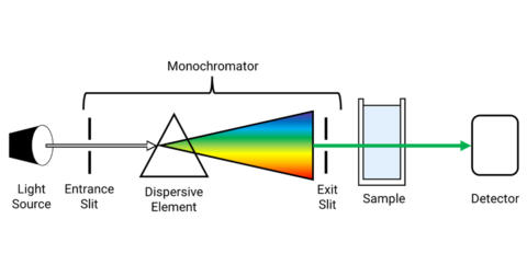
Molecular Spectroscopy
Lab Equipment
Ultraviolet Products
Hybridization Ovens
UVP Incubator
UV Crosslinkers
UVP Benchtop Transilluminators
Thermal Mixers
Electrophoresis & Blotting
Thermostats
View All
Fume hood
Laminar Airflow
Biosafety Cabinet
Autoclave
Centrifuge
pH Meter
Shaker & Mixer
Orbital Shaking Incubator
BOD Incubator
Heating Oven
Water Purification System
Aermax - Air Purification
Medical Oxygen Concetrators
Hygiene Solution
-150°C Cryogenic Freezer
-86°C Ultra Low Temp Freezer
-40°C Low Temp Freezer
-18 ~ -25°C Biomedical Freezer
-20°C Biomedical Freezer
4° ± 1°C Blood Bank Refrigerators
2~8°C Pharma Refrigerators
2~8°C ICE Lined Refrigerators
-25°C ~ + 4°C Mobile Freezer/Collers
20~24°C Blood Platelet Incubators
Ice Machines
Coldrooms
Mortuary Chambers
When the beam of light is passed through the prism and strikes the metal surface like gold and silver, it generates electron charge density waves at the inference between two media called plasmons. The light passed through the metal surface gets reflected with reduced intensity.
The angle at which surface plasmons occur is known as the resonance angle. SPR phenomenon results in the graded reduction of the intensity of light depending on the thickness of a molecular layer at a metal surface.
Surface Plasmon Resonance (SPR) enables label-free detection of biomolecular interactions in real-time. The applications of SPR primarily involve life science research, quality control, electrochemistry, gas phase, and pharmaceutical development.
SPR nature of sensitivity to the refractive index of the medium next to the metal surface has resulted in many biomedical applications. This nature of SPR makes it possible to measure the absorption of molecules and their interactions with specific ligands accurately.
In recent days, the SPR technique is widely used in biomedical applications. This technique is used for various purposes such as real-time measurement of the kinetics of ligand-receptor interactions, screening of compounds in the pharmaceutical industry, enzyme-substrate interactions, epitope mapping, measurement of DNA hybridization, polyclonal antibody characterization, protein conformation studies, and label-free immunoassays.
In the year between 1902 to 1912 R.W. Wood at John Hopkin University (Baltimore, USA) observed that when the polarized light hits the metal-backed diffraction grating, dark light bands appeared in the reflected light. His research was about light, gratings, and metal interactions but there is no answer for this phenomenon was provided.
In 1907 Lord rayleigh based his theoretical research on the scattering of light in the electromagnetic field. He found that the scattered electromagnetic field was singular at wavelengths for one of the spectral orders that arose from the grating at the grazing angle. These wavelengths λR are said to be rayleigh wavelengths that correspond to wood anomalies.
It is observed that these singularities appeared only when the rulings are perpendicular to that polarized electric field and are accounted for S anomalies for P polarization. In the later years, Palmer observed that both the P anomalies and S anomalies are obtainable but the P anomalies were found only in the deep ruled gratings.
In 1941 theoretical analysis undertaken by Fano concluded that these anomalies were related to surface waves or surface plasmons supported by the grating.
In 1968 Otto demonstrated Surface Plasmon Resonance but it was made commercially available for biomolecular interaction after 1990.
Surface Plasmon Resonance occurs when the polarized light hits the metal surface (typically gold or silver) at the interface of media with different refractive indices. SPR techniques excite and detect the oscillations of free electrons on the metal surface that is termed surface plasmons. With Kretschmann–Raether configuration, the light is focussed on the metal surface through the prism and the reflection of light is detected.
At a certain incident angle or resonant angle, the metal surface absorbs light as the plasmons are set to resonate. This results in a dip in reflection intensity and creates a dark band in the detector as shown below -
Most frequently proteins are immobilized on the metal surface, potential ligands are injected over the surface through a series of flow cells. During these biomolecular interactions, the resonant angle of minimum reflected light intensity is detected.
This resonant angle changes as the molecules bind (association constant, Ka) and the dissociation (dissociation constant, Kd). These interactions are recorded in real-time with the SPR sensorgram.
SPR is a common technique with some variations are described above
In case of BLI, sensor tips are used and is dipped into the sample during measurement rather than flowed over the surface as in SPR.
Conclusion
Surface Plasmon Resonance is a phenomenon that is widely used in recent days. This technique is applied in biosensing instruments extensively used to determine the association and dissociation kinetics, nucleic acid hybridization, protein-ligand, protein-protein interactions. SPR spectroscopy is important to carry out nanostructure, and also in label-free chemical and biological sensing.