Sample Preparation
Homogenization
Heating and Mixing
Electrophoresis and Blotting
Polyacrylamide Gel Electrophoresis
Agarose Gel Electrophoresis
Western Blotting
Power Supplies
PCR & qPCR Thermal Cycler
Thermal Cycler (PCR)
Real-time Thermal Cycler (qPCR)
PCR Workstations & Cabinets
UVP BioImaging Systems
Molecular Spectroscopy
Lab Equipment
Ultraviolet Products
Hybridization Ovens
UVP Incubator
UV Crosslinkers
UVP Benchtop Transilluminators
Thermal Mixers
Electrophoresis & Blotting
Thermostats
View All
Fume hood
Laminar Airflow
Biosafety Cabinet
Autoclave
Centrifuge
pH Meter
Shaker & Mixer
Orbital Shaking Incubator
BOD Incubator
Heating Oven
Water Purification System
Aermax - Air Purification
Medical Oxygen Concetrators
Hygiene Solution
-150°C Cryogenic Freezer
-86°C Ultra Low Temp Freezer
-40°C Low Temp Freezer
-18 ~ -25°C Biomedical Freezer
-20°C Biomedical Freezer
4° ± 1°C Blood Bank Refrigerators
2~8°C Pharma Refrigerators
2~8°C ICE Lined Refrigerators
-25°C ~ + 4°C Mobile Freezer/Collers
20~24°C Blood Platelet Incubators
Ice Machines
Coldrooms
Mortuary Chambers
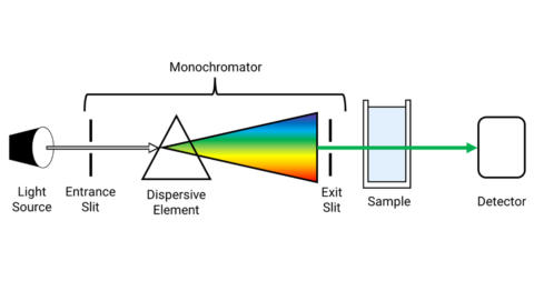
UV/VIS spectrophotometer work on the principle of electromagnetic radiation from the light source to the sample. Depending on the spectrophotometer design set up the light is transmitted through the sample and the light absorbed by the sample is detected using detectors.
In general, a spectrophotometer consists of four main components.
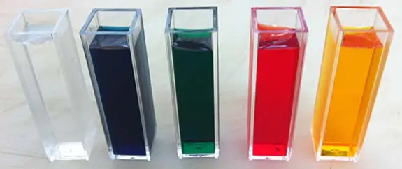
– In the case of liquid samples, Cuvettes are made of quartz, borosilicate glass. Glass and acrylic plastic will never transmit UV light and are used for measurements in the visible range.
– In the case of solid samples, a suitable holder is positioned in the optical path of the spectrophotometer for the measurement of transmitted light.
UV Vis spectroscopy can be classified by the geometry of the components and the optical system. Generally, there are two configurations used in UV Vis spectroscopy are,
The Scanning spectrophotometer works on the principle of measurement of transmittance value at each wavelength. The light is first diffracted with the dispersion element such as quartz, prism, or diffraction grating. The grating is then rotated to select the individual wavelength to send through a cuvette.
The light is then transmitted through the cuvette is recorded. The spectrum that is obtained through the sample solution is continuously observed while changing the wavelength of the light by rotating the grating.
In a scanning spectrophotometer, the grating is mechanically rotated by the motor and so the time taken for the full spectrum is more. Depending on the scanning speed, the scanning process may lead to a decrease in accuracy and reproducibility of the wavelength selection.
In an array spectrophotometer, all the spectral components of the UV Visible range are passed through the sample. The sample in the cuvette through which the light is passed absorbs light at different wavelengths. The light transmitted through the sample is then diffracted by a reflection grating.
This configuration is also known as “ reverse optics” since the light is diffracted with gratings only after passing through the sample. The diffracted light which comprises the light of different wavelengths is detected using sensors and recorded. Generally detector such as long array photosensitive semiconductor material allows simultaneous measurement of all wavelengths of the transmitted light.
This setup allows measuring the full spectrum faster compared to that of a scanning spectrophotometer as the spectrum is measured simultaneously at all wavelengths. The photo array detector used in the array spectrophotometer has an integrating function that accumulates individual measurements to enhance the signal which in turn strongly increases the signal-to-noise ratio. This provides the improved signal quality of the measured spectrum.
Thus the array spectrophotometers with reverse optics technology provide a robust design without any moving optical parts ensures better optical performance with fast measurement of full-spectrum and enhanced quality output.
An optical pathway is a path through which a light beam from the light source is passed through the sample to reach the detector. UV/Vis spectrophotometer has two different pathways either single beam or double beam.
Single beam spectrophotometer has a simple and easiest setup. Here the light beam is directly passed through the sample and to the detector. In order to determine the blank value, a cuvette containing only solvent has to be measured. After determining the blank value, the solvent cuvette is replaced by the cuvette containing the sample to get the absorption spectrum of the sample.
In the double beam spectrophotometer, the light beam is split into a reference and sample beam. There are two different options are available in this optical pathway.
Simultaneous in time – The light beam from the light source are split into two beams of equal intensities and passed through two different cuvettes. One is the reference cuvette and the other is the cuvette that contains sample solution. The light intensity of both beams is measured simultaneously with two detectors.
Alternating in time – Optical Chopper (OC) which is a rotating sectional mirror with which the light is directed alternately through a sample and reference cell. In such a case a unique detector is used to measure both light beams one after the other.
Read more : Difference Between Single Beam And Double Beam Spectrophotometer
Generally, the light source consists of xenon lamp or tungsten and deuterium used for ultraviolet (UV) as well as visible and near-infrared wavelengths over a spectral range from 190nm to1100nm. The light source is directed towards a glass fiber which drives the beam of light onto a cuvette that contains sample solution. The beam passed through the sample absorbs specific wavelengths.
The light that is transmitted through the cuvette is then passed through the diffraction grating that separates light into different wavelengths and detectors such as Photo Diode Array (PDA) or silicon photodiodes, or Charge-Coupled Devices (CCD) is used to record the spectra. As the whole spectrum is measured simultaneously helps in fast recording.
Read more : UV-Vis Measurements – Selecting the Optimum Parameters
Micro-volume UV/VIS spectroscopy
A Micro-volume spectrophotometer is capable of measuring very small volumes and highly concentrated samples. The sample without any dilution is pipetted in the measuring platform. This enables to avoid manipulation errors.
The selection of specific path length enables a high concentration range with a small volume of 1 µL of the sample. The Microvolume spectrophotometer has both a micro-volume platform and a cuvette holder. Depending on the application the light source is flashed either on the micro-volume or cuvette holder.
Conclusion
Emergence and enhancement in power electronic devices have resulted in the robust design of spectrophotometers. Array spectrophotometer is faster compared to that of traditional scanning spectrophotometer.
The single-beam spectrophotometer is cost-effective but if the reference and sample are to be measured simultaneously then the double beam spectrophotometer is chosen.
Depending on the requirement of the lab, the spectrophotometer design is chosen. If there is a requirement to determine the concentration for the low volume samples then micro-volume spectrophotometers are used.