Sample Preparation
Homogenization
Heating and Mixing
Electrophoresis and Blotting
Polyacrylamide Gel Electrophoresis
Agarose Gel Electrophoresis
Western Blotting
Power Supplies
PCR & qPCR Thermal Cycler
Thermal Cycler (PCR)
Real-time Thermal Cycler (qPCR)
PCR Workstations & Cabinets
UVP BioImaging Systems
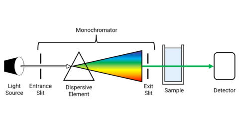
Molecular Spectroscopy
Lab Equipment
Ultraviolet Products
Hybridization Ovens
UVP Incubator
UV Crosslinkers
UVP Benchtop Transilluminators
Thermal Mixers
Electrophoresis & Blotting
Thermostats
View All
Fume hood
Laminar Airflow
Biosafety Cabinet
Autoclave
Centrifuge
pH Meter
Shaker & Mixer
Orbital Shaking Incubator
BOD Incubator
Heating Oven
Water Purification System
Aermax - Air Purification
Medical Oxygen Concetrators
Hygiene Solution
-150°C Cryogenic Freezer
-86°C Ultra Low Temp Freezer
-40°C Low Temp Freezer
-18 ~ -25°C Biomedical Freezer
-20°C Biomedical Freezer
4° ± 1°C Blood Bank Refrigerators
2~8°C Pharma Refrigerators
2~8°C ICE Lined Refrigerators
-25°C ~ + 4°C Mobile Freezer/Collers
20~24°C Blood Platelet Incubators
Ice Machines
Coldrooms
Mortuary Chambers
When electromagnetic energy undergoes interaction with a material, it can be absorbed, reflected, scattered, or transmitted. One kind of scattering is Raman scattering, which serves as the base for Raman spectroscopy. Raman scattered light provides data of the vibrational modes of the samples due to which Raman spectroscopy is beneficial, offering Raman spectrum. Similar to IR spectroscopy, Raman spectroscopy imaging entitles the identification of molecules and their functional groups. This article deals with a variant of Raman spectroscopy, which is micro Raman spectroscopy. Information and science about micro Raman spectroscopy have been briefed.
In micro Raman spectroscopy, a Raman microspectrometer is employed in the place of a conventional Raman spectrometer. A Raman microspectrometer contains a specifically designed Raman spectrometer, which is amalgamated with an optical microscope. This facility permits the technician to acquire Raman spectra of microscopic samples and microscopic areas of macroscopic samples.
The pros of using micro Raman spectroscopy are:
For obtaining different information about the sample from the Raman scattering, micro Raman spectrometer is able to be configured with different colored lasers.
A Micro Raman spectrometer is also referred to as a Raman microspectrometer. It combines both the aspects of Raman spectrometer and microscope. It is purposely built equipment that can do Raman spectroscopy and digital imaging.
A microscope, when used individually, is optical equipment that uses mirrors and lens for the production of magnified images of microscopic objects or microscopic areas of macroscopic objects. The sample substance is illuminated through the objective, with a laser consisting of a very narrow wavelength range.
Gathering the Raman scattered light from the sample is the objective of the microscope. The light is concentrated and frames an image on the entrance aperture of the Raman spectrometer.
The Raman spectrometer portion is a kind of optical instrument utilized for the estimation of the intensity of light relative to its Stokes shift from the wavelength of the exciting laser light. The shit is provided in wavenumbers. A beam of light is collected from a sample, which enters the device, and diffraction grating separates the Stokes shifted frequencies. The separated light is concentrated onto a CCD array detector, in which the intensity of each frequency is estimated by an individual pixel on the array.
Then, the CCD is read-off to a computer monitor and the result obtained is a spectrum, which reveals the intensity of inelastically scattered light versus wavenumbers in relation to the wavelength number of the laser used for excitation.
In a micro Raman spectrometer, a Raman spectrometer is incorporated with a specially designed microscope. The Raman spectrometer is built into a microscope with a digital imaging system, which gives rise to the collection of the maximum amount of light from the smallest samples. In addition, they are very flexible instruments for the measurement of Raman spectra of microscopic areas.
Micro Raman spectroscopy is used for the identification of different molecules and functional groups present within larger molecules. Bonds formed between the atoms possess specific vibrational frequencies, which correspond to the masses of atoms and the potency of the bond between them. The complex molecules display many peaks and can be promptly recognized by the fingerprint or pattern created by the peaks. There are plenty of applications of micro Raman spectrometers as they are able to recognize microscopic samples or microscopic areas of bigger samples in a non-destructive way.
Forensics
Semiconductor
Art
Geology
Materials Science
Pharmaceutical
Miscellaneous
Micro Raman spectroscopy is perceived as a conventional Raman technique merely with superior resolution, which is bounded by a far-field diffraction limit.
Micro Raman spectroscopy is also known as Raman microscopy. It embraces the pairing of a conventional Raman spectrometer with an optical microscope. This conjugation delivers immense power in sampling portions with higher spatial resolution in the order of 1 micrometer.
Types of Lasers used
For a Raman microscopy set up, the following laser sources can be used:
For obtaining reliable and precise results of the sample, the calibration of the wavelength axis is a requisite. Several operational changes in Raman microscopy generally possess less or more severe consequences in terms of calibration of wavelength. Though modern microscopes provide continuous calibration for maximum convenience, recalibration is done by estimating a silicon standard. If the equipment is not calibrated continuously, recalibration should be done routinely. Minor adjustments like aperture, grating, mirror, vibrations, sudden shocks, variations, temperature shifts are performed to ensure delivery of optimal spectral data.
Conclusion
Initially, a short introduction to Raman spectroscopy and micro Raman spectroscopy has been provided. The pros of micro Raman spectroscopy have been discussed. A way of designing a micro Raman spectrometer has been given. Then, applications of micro Raman spectroscopy in different fields have been listed. Laser usage in Raman microscopy has been explained. Subsequently, calibration of Raman microscopy has been detailed.