Sample Preparation
Homogenization
Heating and Mixing
Electrophoresis and Blotting
Polyacrylamide Gel Electrophoresis
Agarose Gel Electrophoresis
Western Blotting
Power Supplies
PCR & qPCR Thermal Cycler
Thermal Cycler (PCR)
Real-time Thermal Cycler (qPCR)
PCR Workstations & Cabinets
UVP BioImaging Systems
Molecular Spectroscopy
Lab Equipment
Ultraviolet Products
Hybridization Ovens
UVP Incubator
UV Crosslinkers
UVP Benchtop Transilluminators
Thermal Mixers
Electrophoresis & Blotting
Thermostats
View All
Fume hood
Laminar Airflow
Biosafety Cabinet
Autoclave
Centrifuge
pH Meter
Shaker & Mixer
Orbital Shaking Incubator
BOD Incubator
Heating Oven
Water Purification System
Aermax - Air Purification
Medical Oxygen Concetrators
Hygiene Solution
-150°C Cryogenic Freezer
-86°C Ultra Low Temp Freezer
-40°C Low Temp Freezer
-18 ~ -25°C Biomedical Freezer
-20°C Biomedical Freezer
4° ± 1°C Blood Bank Refrigerators
2~8°C Pharma Refrigerators
2~8°C ICE Lined Refrigerators
-25°C ~ + 4°C Mobile Freezer/Collers
20~24°C Blood Platelet Incubators
Ice Machines
Coldrooms
Mortuary Chambers
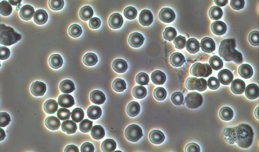
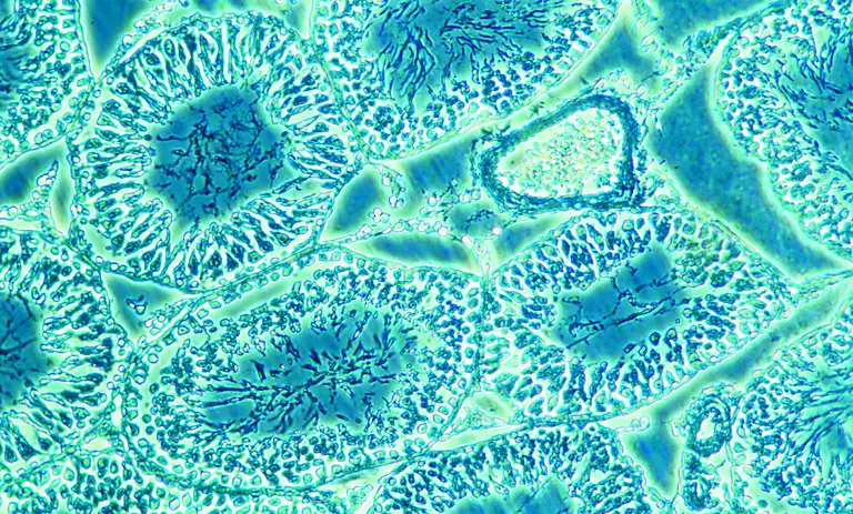
Translucent biological specimens are hard to get observed in a traditional conventional microscope. To solve this problem, researchers and scientists use phase-contrast microscopy techniques. With the help of this microscopy technique, one can gain images of biological samples in high contrast without staining. Phase-contrast microscopy is a famous technique that is used for unstained biological samples. Its high-resolution objective lens enables the formation of high-contrast images that gives a detailed structure of specimens under the microscope. All components of the phase-contrast microscope are well designed to form the above-mentioned images. This category of the microscope is also called a cell culture microscope, tissue culture microscope, and culture microscope.
Several types of biological specimens can be thin or thick. Some biological samples are translucent which do not absorb or reflect the incident light rays much. In this case, the diffraction and reflection happen in the phase-contrast microscopy technique that helps in the generation of high contrast images. The special condensers are used in the inverted phase-contrast microscope that mediates the light rays from the light source to get interfered. Zernike 1932 formulated the phase-contrast microscope in his university with the help of his colleagues. From that time, the inverted phase microscope was used for various clinical aspects and research. In the diagnostics area also, the trinocular compound microscope of phase-contrast nature can be used.
As per this technique, the phase-contrast microscope exploits the electromagnetic nature of light rays. The light rays steadily pass through a biological specimen over its plane. Here, the biological sample is placed on the pointing stage of the phase-contrast microscope. When light rays fall on a biological sample, the whole of it gets illuminated by a hollow cone of light. The phase rings present in the optical device mediates the light rays to the specimen through the condenser.
The phase-contrast microscopy technique should be carried out with an efficient phase-contrast objective lens. Each of the annulus rings should be used with the corresponding phase objective lens. An annulus ring can be located in the part of the trinocular head. In this trajectory, the light rays get a little retarded from their usual path length. Further, retardation takes place in the phase plates used up in the trinocular microscope. All light rays then combine to cause the interference process. It is seen that in the inverted phase-contrast microscope, the light rays which have not taken part in any destructive and constructive interferences, produce a characteristic light on the specimen. These light rays are responsible for the generation of high contrast images in phase-contrast microscopy. Here, the light rays also form the dark images of the biological samples used in phase-contrast microscopy. The specimens used in phase contrast microscopy should be small enough to get the magnified image in the microscope. Mounting on the gel can also happen in the specimen for a better bioimaging process. The best trinocular microscope possesses the feature of phase-contrast microscopy for better magnification of all biological specimens. It is all used for many research as well as diagnostics areas.
It is well observed that a phase-contrast microscopy technique utilizes a special way for producing enlarged images. Due to this special technique, it is used in different areas of both clinical well as research areas. Many hospitals and research centers are using phase-contrast microscopy techniques for concluding their work. All transparent biological specimens get magnified with this imaging technique. In a brightfield microscope, the transparent specimens cannot be observed closely. On the contrary, the phase-contrast microscope can very efficiently produce images of all biological specimens with good resolutions.
Following are some of the advantages of phase contrast microscopy in clinical settings :
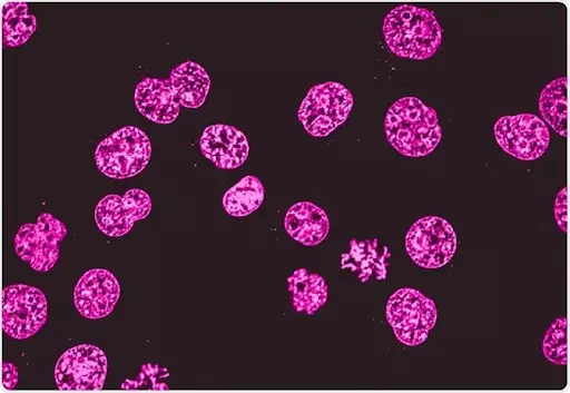
In the early days of phase-contrast microscopy, scientists were unable to observe the living cells. Frits Zernike has made the observation of all living cells very easy with his discovery. In the absence of the phase-contrast microscopy technique, the cells were dried and then mounted on the slides for observations. This made room for losing the vitals of the cells. But with a phase-contrast microscope, the researchers can directly observe the living cells without the staining process.
Light rays making a different contrast in the view of all biological specimens make the observations of living cells easy. The cells become more illuminated and could be easily studied with the help of an inverted phase microscope.
Biological specimens which are transparent and translucent can be viewed very minutely in phase-contrast microscopy. For example, a very thin slice of tissue can be viewed perfectly in a phase-contrast microscope. The structures as well as the details all are perfectly seen in the phase-contrast microscopy techniques which are absent in brightfield microscopes. With a phase-contrast image, one can easily view and study the details of the internal structure of a cell or microorganisms, or biological specimens. The contrast can also be adjusted in the microscope for better imaging of all samples.
To get the best-contrasted image for the corresponding biological specimens, scientists or researchers sometimes need to experiment with different observation methods. Phase-contrast microscopy techniques can be easily combined with other methods of magnification, such as fluorescence, etc. The combination reveals additional detail that couldn’t be observed with just one observational method. This combination backs the phase microscopy technique more in clinical settings. For example, fluorophores are used for generating labels for structures of cells. With the phase-contrast microscopy technique, fluorescence can also be used for studying the cells.
Inverted phase contrast microscope or phase contrast microscope is not limited to clinical applications only. Many different applications also prevail in the scientific world. Many types of biological specimens can be easily viewed with a phase-contrast microscope using its interference process. Diffraction and reflection processes are also employed in the phase microscopy technique. Other Applications are as follows :
All Phase contrast microscopes produce a unique “halo” around the edges of samples. This is exclusive of phase contrast microscopy which helps in observing cells. It is particularly useful to rapidly locate tiny specimens or samples in the culture such as bacteria or debris.
Conclusion
Phase contrast microscopy has many applications in the scientific world. In clinical settings also, it works very efficiently, it gives out perfect contrasted images which makes up the best observations for cell cultures and structures of cells. As a lab fellow, you can check the trinocular microscope price online as well as offline medium.