Sample Preparation
Homogenization
Heating and Mixing
Electrophoresis and Blotting
Polyacrylamide Gel Electrophoresis
Agarose Gel Electrophoresis
Western Blotting
Power Supplies
PCR & qPCR Thermal Cycler
Thermal Cycler (PCR)
Real-time Thermal Cycler (qPCR)
PCR Workstations & Cabinets
UVP BioImaging Systems
Molecular Spectroscopy
Lab Equipment
Ultraviolet Products
Hybridization Ovens
UVP Incubator
UV Crosslinkers
UVP Benchtop Transilluminators
Thermal Mixers
Electrophoresis & Blotting
Thermostats
View All
Fume hood
Laminar Airflow
Biosafety Cabinet
Autoclave
Centrifuge
pH Meter
Shaker & Mixer
Orbital Shaking Incubator
BOD Incubator
Heating Oven
Water Purification System
Aermax - Air Purification
Medical Oxygen Concetrators
Hygiene Solution
-150°C Cryogenic Freezer
-86°C Ultra Low Temp Freezer
-40°C Low Temp Freezer
-18 ~ -25°C Biomedical Freezer
-20°C Biomedical Freezer
4° ± 1°C Blood Bank Refrigerators
2~8°C Pharma Refrigerators
2~8°C ICE Lined Refrigerators
-25°C ~ + 4°C Mobile Freezer/Collers
20~24°C Blood Platelet Incubators
Ice Machines
Coldrooms
Mortuary Chambers
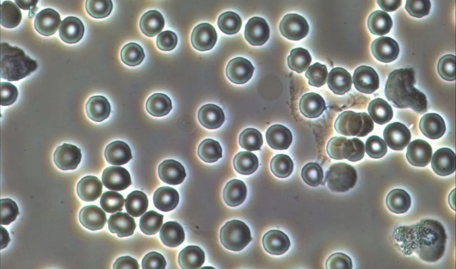
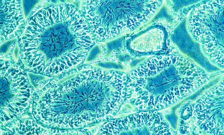
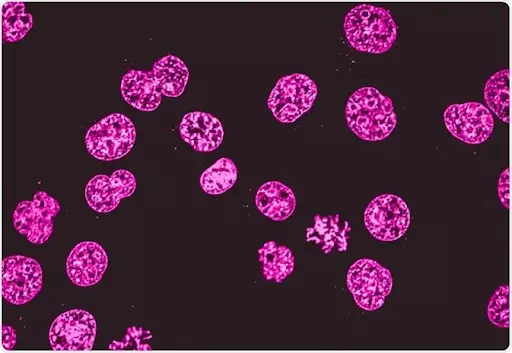
In a research lab, microscopes play a crucial role in observing the structural details of biological samples and molecules. There are different types of microscopy techniques prevalent in the scientific world, which makes the observations of various specimens. One such microscopy technique is phase-contrast microscopy. Under this microscopy, the specimen’s ability to alter the optical light is manipulated. As the direct light penetrates the specimen’ wall, the light gets diffracted through interferences of light. This whole process, then, results in the high contrast image of the specimen under the microscope. A phase-contrast microscope has a trinocular head which helps in the observation process. The different levels of contrast also help in the observation process to make the image more illuminated.
Phase contrast microscopes are also called tissue culture microscopes, because of their contrasting ability to illuminate the image. When the direct source of light is made to pour into the biological sample, the light starts to get diffracted. This diffraction produces the interferences of light which are not that subtle to be seen with naked eyes. While scattering the light or diffracting the light, some of the light gets absorbed by the specimen illuminating it with full contrast. However, the light which is not diffracted in the phase-contrast microscopy is called zero-order light. Here, the zero-order light remains unmodified throughout the magnification process.
The discovery of phase contrast microscopy was made by a Dutch physicist, Frits Zernike in 1938. Phase-contrast microscopy discovery has led Zernike to the prestigious Nobel prize in physics (1953) And after this feat, the Germany-based company called Zeiss started manufacturing the inverted phase-contrast microscope in their labs, during world war II.
Frits demonstrated with the speed of light path and directed towards the specimen. He went on experimenting with optical light paths to discover interference patterns of light. This results into the images appearing darker under the inverted phase microscope. Zernike’s approach consisted of simple and reasonable components. This includes:
The annulus and ring ultimately reduce the light wavelength by a ½ phase. This paves the way for the magnification of biological specimens. To obtain an image of 10x and 100x, the annulus is set into the light condenser of a trinocular compound microscope. A culture microscope also uses the rings for illuminating the images to their threshold magnification.
To obtain the perfect enlarged image of the biological samples, the inverted phase contrast microscope uses some basic components. These components work in harmony to give out the perfect high contrasted image of the specimens.
Following are the basic components of phase contrast microscopy :
The unstained, transparent, and colorless biological specimens are called phase objects. These objects do not absorb direct light. But the biological samples are well good at diffracting the direct light to produce the magnified images. Through the trinocular head, the phase-contrast microscopy process can be observed.
The annular diaphragm is situated below the condenser in a cell culture microscope. It is made up of a circular disc having an annular groove for light passing through the trinocular head of a phase-contrast microscope. As the light rays reach the annular groove of the annular diaphragm, all light rays fall onto the biological specimen. At the backplane, the objective aperture develops the image of a biological sample.
The phase plates used up in an inverted phase-contrast microscope can be either negative or positive. Negative phase plates have a thick circular area that drives the light paths. While the positive phase plates have a thin circular groove to drive light paths. Phase plates come with a transparent disc for rotating them. The phase plates and the annular diaphragm, both work closely to give out the contrasted image of the specimen. The image is obtained by separating the direct light rays from the diffracted ones. All direct light rays fall on the annular groove and the diffracted light rays fall on the region outside the groove. Different refractive indexes give out different images for the inverted phase-contrast microscope.
After the phase plates and phase annulus, the condensers also play a vital role in resulting in contrasted images of the specimen. The phase condensers are well structured for passing the light rays from the phase rings and plates. All diffracted light rays reach the condensers from where it reaches the objective side. The wavefronts from the diffracted light rays collect at one point and produce contrasted specimens. Here, the interference of light waves happens which is the main reason to form the contrasted images of all biological samples.
A light source is very important for the phase-contrast microscopy process. Without the light source, the phase microscope cannot be used. A reliable light source should be used in phase-contrast microscopy for forming the enlarged image of biological samples. Natural sunlight can also be used in the process. However, another artificial light source can also be used.
Following are the advantages of phase contrast microscopy :
Following are the limitations of phase contrast microscopy:
Conclusion
Phase-contrast microscopy is a technique that manipulates the traditional brightfield microscope working mechanism. When all the components of phase contrast microscopy are configured properly, it visualizes the images of the specimen very vividly. The high contrasted images can be obtained with the implantation of the phase contrast microscopy process. Phase imaging eliminates the use of labels in the biological samples. Hence, it saves time for researchers and scientists. Though the phase microscopes are expensive, the trinocular microscope price can be gathered from the online medium as well as offline medium.