Sample Preparation
Homogenization
Heating and Mixing
Electrophoresis and Blotting
Polyacrylamide Gel Electrophoresis
Agarose Gel Electrophoresis
Western Blotting
Power Supplies
PCR & qPCR Thermal Cycler
Thermal Cycler (PCR)
Real-time Thermal Cycler (qPCR)
PCR Workstations & Cabinets
UVP BioImaging Systems
Molecular Spectroscopy
Lab Equipment
Ultraviolet Products
Hybridization Ovens
UVP Incubator
UV Crosslinkers
UVP Benchtop Transilluminators
Thermal Mixers
Electrophoresis & Blotting
Thermostats
View All
Fume hood
Laminar Airflow
Biosafety Cabinet
Autoclave
Centrifuge
pH Meter
Shaker & Mixer
Orbital Shaking Incubator
BOD Incubator
Heating Oven
Water Purification System
Aermax - Air Purification
Medical Oxygen Concetrators
Hygiene Solution
-150°C Cryogenic Freezer
-86°C Ultra Low Temp Freezer
-40°C Low Temp Freezer
-18 ~ -25°C Biomedical Freezer
-20°C Biomedical Freezer
4° ± 1°C Blood Bank Refrigerators
2~8°C Pharma Refrigerators
2~8°C ICE Lined Refrigerators
-25°C ~ + 4°C Mobile Freezer/Collers
20~24°C Blood Platelet Incubators
Ice Machines
Coldrooms
Mortuary Chambers
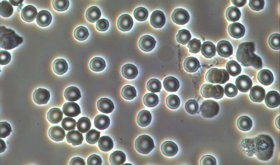
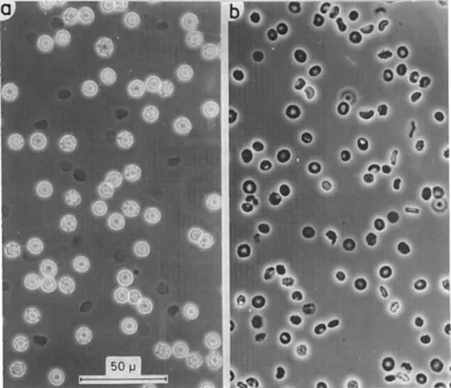
Phase-contrast microscopy falls in the category of light microscopy that offers powerful label-free imaging of biological molecules. These biological molecules can be physical as well as soft materials. With a good trinocular microscope, you can get the right images of the desired biological molecules. The standard light microscope has some limitations of low contrast that does not give out a perfect image of the biological samples. To eradicate this limitation, phase-contrast microscopy is used. Here, the biological sample gets illuminated with good contrast and light. The samples used here, tend to execute the diffraction process which gives out the changes in the various refractive indexes which reveal many structures of the biological samples. However, in some cases, the refractive index differences are so slim that the structures cannot be revealed. But with a perfect inverted phase-contrast microscope, the images can be formed in the microscope’s objective area.
The discovery of the phase-contrast microscope led the discoverer Frits Zernike to the Nobel prize. Phase-contrast microscopy is defined as the process that converts phase shifts of light thrown to the samples to the light entering the transparent sample. Hence, the whole sample gets illuminated with the light entering it. The high contrast label-free images are obtained at the end of the phase-contrast microscopy. The best trinocular microscope of phase-contrast nature is present in the market that can be used to give out the perfect phase-contrast images. Here, the phase shifts are not visible, unless their trajectories are recorded in the form of images. The trinocular microscope is used in this reference. The use of inverted microscope is to give out perfect images.
A phase-contrast microscope does not record the phase variations at a time. But can certainly give out the results in the form of high contrast images. The images which are given out with this type of microscope, are the mere results of amplitude changes between the light shifts. This is called amplitude contrast. Generally, the phase variations are not visible. It needs modifications in a tissue culture microscope of phase-contrast nature to give out the perfect illuminations. The adjustments are done in several parameters before getting the phase-contrast images of the biological samples.
A standard phase contrast culture microscope runs on a certain principle which gives out the perfect illuminated images. All transparent unstained biological samples do not absorb light. So, they are called phase objects. When light passes through samples in a phase-contrast microscope with no phase object, there is no significant change in refractive indexes. The optical path length remains the same with no change. Here, the non-diffracted light is referred to as zero-order light as it continues to be unchanged throughout the process unless there are no phase objects.
When the light passes through a transparent area of the biological sample, then small changes in refractive indexes are measured. The phase objects here diffract the light and scatter the light at the same time. These changes in the refractive indexes of the light phases cause changes in the optical path length of the light and result in the illuminated bio-images. A culture microscope uses this principle to get well-structured images of the culture samples.
Here, the diffractions happen as per the thickness of each structure present in the sample. It is seen that the thicker the structure of the biological sample, the greater is the diffraction of the light. In a phase-contrast microscope, the diffracted light represents only a very small part of total light passing through the sample. A large proportion of light passing through the biological sample is lost to the environment or helpful to the illumination process.
In phase-contrast microscopy, the trinocular head is present which enables the light passing. As the diffracted light arrives at the detector of the microscope, it becomes out of phase with the direct light. The main core of phase contrast microscopy is the interference caused by the phase shifts of light. All small -small phase shifts of light created by the light piercing the transparent structures are not capable of creating the interferences between the phase lights. In addition to these, the feature of low absorption also causes negligible amplitude differences between the phase of lights.
To put it in simple words, phase contrast microscopy is a method or process that manipulates the nature of phase objects to introduce additional interferences. These interferences are introduced between the direct and diffracted light phases in the inverted phase microscopes. These additional interferences transform the diffraction differences into brightness or contrast. It all increases the contrast, which can be seen in the non-absorbing samples. By this principle of a phase object, the phase-contrast microscope can give out illuminated images of the biological samples.
An inverted phase microscope can also be used in the process to get enlarged biological images. The phase-contrast microscope components can be fitted in this, to get the illuminated images to a good degree. The trinocular microscope price can be gathered from the offline as well as an online medium.
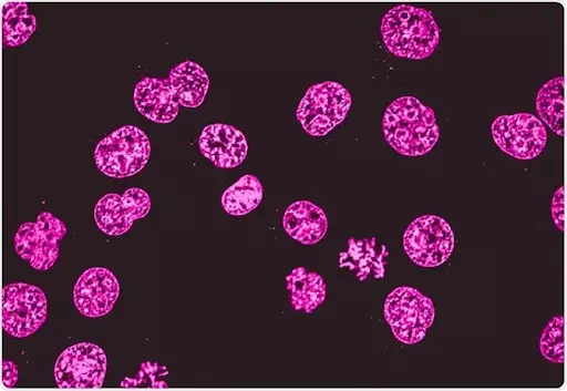
Phase-contrast microscopy is useful in various methods of biological light microscopy processes. In the biological and scientific world, it has a wide variety of applications which are used for cell imaging. Because of the use of a trinocular compound microscope with phase contrast nature in the cell cultures, it is called the culture microscope.
Following are the key areas where the phase-contrast microscopy is used and applied:
Like, these many types of applications are of phase-contrast microscopy. In a biological lab, it finds many applications in the research field.
Conclusion
A phase-contrast microscope is all good for getting an illuminated image of the biological samples. It is all good for observing the small microscopic organelles in full brightness. The thicker the sample, the more light gets diffracted. This ensures that the biological sample works best in the lab for performing the biological research.