Sample Preparation
Homogenization
Heating and Mixing
Electrophoresis and Blotting
Polyacrylamide Gel Electrophoresis
Agarose Gel Electrophoresis
Western Blotting
Power Supplies
PCR & qPCR Thermal Cycler
Thermal Cycler (PCR)
Real-time Thermal Cycler (qPCR)
PCR Workstations & Cabinets
UVP BioImaging Systems
Molecular Spectroscopy
Lab Equipment
Ultraviolet Products
Hybridization Ovens
UVP Incubator
UV Crosslinkers
UVP Benchtop Transilluminators
Thermal Mixers
Electrophoresis & Blotting
Thermostats
View All
Fume hood
Laminar Airflow
Biosafety Cabinet
Autoclave
Centrifuge
pH Meter
Shaker & Mixer
Orbital Shaking Incubator
BOD Incubator
Heating Oven
Water Purification System
Aermax - Air Purification
Medical Oxygen Concetrators
Hygiene Solution
-150°C Cryogenic Freezer
-86°C Ultra Low Temp Freezer
-40°C Low Temp Freezer
-18 ~ -25°C Biomedical Freezer
-20°C Biomedical Freezer
4° ± 1°C Blood Bank Refrigerators
2~8°C Pharma Refrigerators
2~8°C ICE Lined Refrigerators
-25°C ~ + 4°C Mobile Freezer/Collers
20~24°C Blood Platelet Incubators
Ice Machines
Coldrooms
Mortuary Chambers
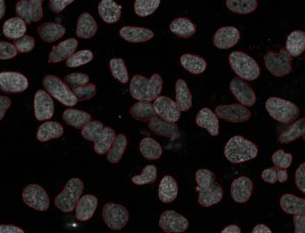
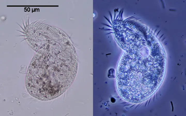
The phase contrast microscopy technique is one of the types of microscopic techniques that are used by scientists and researchers for observing unstained biological specimens. These specimens are called phase objects or flat cells. All these cells are visible under an optical phase contrast microscope through their trinocular head. Phase contrast microscopy visualizes the images of those cells which appeared inconspicuous and transparent in the brightfield and darkfield microscopes. It can be combined with an inverted microscope to make an inverted phase microscope and an inverted phase contrast microscope. These types of microscopes are also called cell culture microscope, culture microscope, and tissue culture microscope. The unstained cells are visualized in high contrast and much detail under the phase contrast microscope. For this reason, it is used in a biological lab mostly.
All biological specimens used up for the phase contrast microscopy are called phase objects. These phase objects cause a shift in phases in the light passing through itself. The images formed in the phase contrast microscope are highly contrasted which is the resultant of the amplitude shifts, both in refractive index differences and intensities. These differences are visible to the human eye or photo detectors for magnifying the images. The staining of the biological specimen would help in mediating this amplitude shift and also help in acing the differences in the intensity of light passing through.
It is seen that many of the staining reagents for living cells are toxic to it. So, the researchers and scientists, use the phase contrast microscope for viewing such objects. In this case, the phase contrast microscopy technique is used that offers the change in phase shifts to help in causing differences in the source light’s optical path. This all makes the path visible and ultimately, helps in illuminating the biological specimen. The microscope changes the phase shifts into amplitude shifts through the interference of resulting all the light rays waves. All these changes can help in the magnification of the biological specimens under a trinocular compound microscope.
This elite technique of phase contrast microscope was developed in the 1930s by Dutch physicist Frits Zernike by combining the simple compound microscope to form the phase differences. For this discovery, he was awarded the Nobel Prize in Physics in 1953.
The phase contrast microscopy technique manipulates the electromagnetic nature of light entering over the surface of phase objects or specimens. Here, the optical path length is called the product of the refractive index. This thickness is between two points in an optical path. All this is related to the transit time and the velocity of light. Here, the differences in optical path length lead to differences in velocities of the source light waves. It passes through the biological specimens. it results in the phase differences in the optical light waves that pass through the specimens. It is seen that a higher refractive index‘s biological specimen compared to the surrounding medium paves the way for the deceleration of the light waves and also causes the retardation of its phase.
In phase-contrast microscopy, interferences describe the interaction of two light waves with each other. This results in the formation of a new wave pattern that follows the principle of superposition. This makes up the relevant parameter for the interference of light waves’ amplitude. In this condition of light interferences, if the two light waves interfere, the amplitude of the resulting light wave becomes equal to the vector sum of the amplitudes of two interfering light waves. The interferences caused here, form the phase contrast images of the biological specimens.
Moreover, if the amplitude of the resulting waves is increased, then the interferences formed will be constructive. In this scenario, it is also seen that the crest of one wave and a trough of another wave meet at the same point in time. This all leads to a decrease in the amplitude of the resulting wave. Here, the interference is called destructive nature. But in the actual microscopy technique, the constructive interferences cause the image formation. This helps in the magnification of the biological specimens.
In a typical inverted phase contrast microscope, the main components are as follows :
Apart from this, condensers and objective lens is also used to visualize the images. In the microscopic setup, the annulus aperture is placed in the front of the focal plane of the condenser which limits the angle of penetrating light waves through the trinocular head. The phase plates in the inverted phase microscope lie in the back focal plane of the objective lens which has a phase ring. This phase ring is made up of material that dims the light passing through it and also changes its phase by λ/4. λ represents the wavelength of light in the phase contrast microscope.
In Kohler’s case of illumination, the light waves which do not interact with each other with the sample, all get focussed as a bright ring in the back focal plane of the objective lens in a microscope. Here, the light rings formed spatially match the phase rings along the optical axis of the optical light length. All these cause a phase shift in the undeviated light rays. The light rays that get diffracted by the biological specimen do not predominantly strike the phase rings on the microscope. They remain non-affected by the light rays.
In between the affected light and unaffected light waves, there is a total phase shift of up to λ/2. Here, the phase of undeviated light is advanced by λ/4 at the phase ring. It gets diffracted by light waves that are usually retarded by λ/4 by biological samples. The total phase shift of λ/2 offers the destructive interference of the light waves in the image plane of the microscope. To dim out the undeviated light rays passing the phase ring, it is important to avoid an outshining of the undeviated light rays compared to the deviated light rays. The best trinocular microscope can illuminate biological samples.
In phase contrast microscopy, the phase shift of λ/2 is observed. It gives rise to a maximal destructive interference effect as crest and trough effectively that meet at the same point in time. Hence, the amplitude of the light wave is reduced and the phase shift of the phase object is transformed into an amplitude shift.
After the formation of destructive and constructive interference phase differences, the highly contrasted image is formed. The phase contrast microscopy technique visualizes differences in the optical path length of a biological specimen. All cellular structures like plasma membranes, different organelles affect the optical path length. The images appear in the form of halo or highly contrasted images of it which is hardly visible in brightfield microscopy. These cells amplify the phase differences in the contrasted images that can be mapped in an optical density map.
The halo describes the appearance of a bright edge of the positive phase contrast or a dark edge for negative phase contrast around the large objects. Halos forms during the phase contrast microscopy technique are because of the diffracted light from the biological specimen.
Conclusion
The phase contrast microscopy technique is a subtle way to form the high contrasted images of all types of biological specimens. The interferences and diffraction of light rays produce the phase changes in the optical light paths which helps in the formation of high contrasted images of unstained biological specimens.