Sample Preparation
Homogenization
Heating and Mixing
Electrophoresis and Blotting
Polyacrylamide Gel Electrophoresis
Agarose Gel Electrophoresis
Western Blotting
Power Supplies
PCR & qPCR Thermal Cycler
Thermal Cycler (PCR)
Real-time Thermal Cycler (qPCR)
PCR Workstations & Cabinets
UVP BioImaging Systems
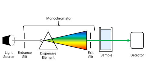
Molecular Spectroscopy
Lab Equipment
Ultraviolet Products
Hybridization Ovens
UVP Incubator
UV Crosslinkers
UVP Benchtop Transilluminators
Thermal Mixers
Electrophoresis & Blotting
Thermostats
View All
Fume hood
Laminar Airflow
Biosafety Cabinet
Autoclave
Centrifuge
pH Meter
Shaker & Mixer
Orbital Shaking Incubator
BOD Incubator
Heating Oven
Water Purification System
Aermax - Air Purification
Medical Oxygen Concetrators
Hygiene Solution
-150°C Cryogenic Freezer
-86°C Ultra Low Temp Freezer
-40°C Low Temp Freezer
-18 ~ -25°C Biomedical Freezer
-20°C Biomedical Freezer
4° ± 1°C Blood Bank Refrigerators
2~8°C Pharma Refrigerators
2~8°C ICE Lined Refrigerators
-25°C ~ + 4°C Mobile Freezer/Collers
20~24°C Blood Platelet Incubators
Ice Machines
Coldrooms
Mortuary Chambers
Raman spectroscopy is one of the non-destructive chemical analysis and spectroscopic techniques, which offers detailed information about polymorphy, crystallinity, phase, chemical structure, and molecular interactions. The Raman spectroscopy is based on the Raman scattering technique, which is due to the interaction of light with the chemical bonds present within a material. When the molecule scatters incident light obtained from a high-intensity laser light source, Raman lines are obtained on the Raman spectrum. With the aid of both spectral and spatial information, Raman imaging is performed and images are generated. Raman analysis is performed using a spectrometer for the identification of the “chemical fingerprint” of the desired molecule. When the incident light beam is deflected by the molecules, there is a change in the wavelength of the light, which is the Raman effect.
Raman spectroscopy takes on the understanding of various systems at molecular levels since its discovery. In JNCASR, two custom-built micro-Raman setups have been fabricated in a laboratory. One setup uses a red laser for studying specifically biomolecular interactions. Another has green-colored laser excitation at 532 nm for probing interactions in inorganic compounds and crystalline-like structures. Also, works are being carried out for setting up a polarization-based Raman spectrometer to detect Raman optical activity in various biomolecules, especially proteins. A high-resolution Raman spectrometer having four different lasers (405 nm, 532 nm, 633 nm, and 785 nm), which are used for SERS, temperature-dependent and pressure-dependent studies, and magnetic-Raman studies. This article focuses on the applications of the fabricated high-resolution Raman spectrometer.
The high-resolution Raman spectrometer is used in the following applications:
Magneto-Raman studies can be performed with magnetic fields up to 0.6 T. and in the temperature ranges of 3K-350 K, which serves exciting scope in the exploration of engaging magnetic properties and phase transitions.
Temperature-dependent Raman studies are useful in the identification of magnetic, electronic, and structural transitions.
Raman scattering is used to capture vibrational modes of materials and molecules. Minute structural changes caused by external stimuli like pressure or temperature are reflected as shifts in the vibrational modes of frequency and changes in intensity. The Raman spectroscopy technique is utilized for the investigation and understanding of phase transitions and gas adsorption in metal-organic frameworks (MOF) like ZIF-7, ZIF-8, and MOF-508.
A sensitive, sturdy, and cost-effective miniature tabletop Raman spectrometer has been developed. The developed Raman spectrometer offers a decent spectral resolution with a high throughput attained by f-number. It is coupled to a large clear aperture for effective collection of light in a custom-made spectrometer. The time of integration ranges from milliseconds to a few seconds, which helps in achieving faster scans. The system has been combined with a motorized stage, where accommodation of 96 well plates is done. It has been automated for running Raman maps along with these wells, to achieve automated multi-well maps for quick scans for greater sets of samples. The demonstration of SERS of R6G is done in a 5ms time, but with similar experimental conditions, a commercial spectrometer can acquire a similar intensity spectrum of the same sample in 4 min.
Pressure is a clean and effective tool for studying material properties as it is one of the fundamental thermodynamic parameters. Pressure can change the force, volume, electronic structure, effective hybridization, interatomic bond distances, density, and accordingly, material intrinsic characteristics. Raman scattering along with synchrotron X-ray diffraction are versatile and efficient probes for studying the structural and electronic transitions provoked by external stimuli like pressure. Interesting phenomena like topological quantum phase transitions, structural phase transitions, amorphization, isostructural transitions, lattice dynamics, and insulator-metal transitions can be examined under extremely high-pressure conditions using this technique.
Raman scattering and synchrotron XRD techniques are used to understand the electronic and structural phase transitions in condensed matter systems induced by pressure. Raman scattering has been utilized in probing structural distortions of a few of the rare-earth functional oxides and electronic transitions of high spin-orbit coupling materials as a function of pressure. Recently, study and identification of topological quantum phase transitions in materials like 1T-TiTe2 and 1T-SeTe2.
Raman spectroscopy, though a weak phenomenon, is a powerful tool. Nanostructures synthesized from plasmonic metals like silver, copper, and gold offer an enhanced electric field in their vicinity during the excitation of electric fields due to the phenomenon of localized surface plasmon resonance (LSPR). Biomolecules are generally weak Raman scatterers and in the presence of plasmonic nanostructures, their spectra get highly increased.
Aurora kinase enzymes are relative to tumorigenesis and are strong anti-cancer drugs. The orientation of the enzyme on the surface of silver nanoparticles can result in a different Raman spectrum. Felodipine is a specific and allosteric inhibitor for the enzyme oncogenic Aurora kinase A, which is confirmed through analysis of the SERS spectrum of Aurora kinase A and in the presence of a molecule.
Extracellular vesicles (EVs) laden with DNA, micro-RNA, proteins, and lipids play significant biological functions in intracellular communication and possess essential roles in pathophysiological conditions. Simple citrate reduced silver nanoparticles aided SERS as a tool to differentiate EVs extracted from various cell lines like Atg5 and HeLa isolated under autophagic conditions (nitrogen starvation). The nanoparticles aided in the facile differentiation and detection of EVs isolated between two closely related human cells, which differ in their autophagic capability. The existence of distinct chemical compositions of EVs is constantly revealed by the principal component analysis (PCA) of SERS Raman spectra of EVs.
HIV-I is categorized into 9 distinct and primary genetic subtypes designated A to J (E and I are recombinants). The distribution of subtypes of the virus across the globe is non-uniform. Through SERS and resonance Raman spectroscopy (SERRS), capture probes for capturing nucleic acids have been discovered for development of a high sensitivity assay for molecular typing of a biological sample. Along with a kit for molecular typing of biological samples using SERS, detector probes are designed for the detection of capture probes. Through the assay, detection of HIV RNA is possible. In addition, it also differentiates the various subtypes of HIV-1.
Conclusion
A brief introduction to Raman spectroscopy and related terms have been provided. A short description of the development of a high-resolution Raman spectrometer has been given. Finally, Five major applications of the high-resolution Raman spectrometer have been discussed and detailed.