Sample Preparation
Homogenization
Heating and Mixing
Electrophoresis and Blotting
Polyacrylamide Gel Electrophoresis
Agarose Gel Electrophoresis
Western Blotting
Power Supplies
PCR & qPCR Thermal Cycler
Thermal Cycler (PCR)
Real-time Thermal Cycler (qPCR)
PCR Workstations & Cabinets
UVP BioImaging Systems
Molecular Spectroscopy
Lab Equipment
Ultraviolet Products
Hybridization Ovens
UVP Incubator
UV Crosslinkers
UVP Benchtop Transilluminators
Thermal Mixers
Electrophoresis & Blotting
Thermostats
View All
Fume hood
Laminar Airflow
Biosafety Cabinet
Autoclave
Centrifuge
pH Meter
Shaker & Mixer
Orbital Shaking Incubator
BOD Incubator
Heating Oven
Water Purification System
Aermax - Air Purification
Medical Oxygen Concetrators
Hygiene Solution
-150°C Cryogenic Freezer
-86°C Ultra Low Temp Freezer
-40°C Low Temp Freezer
-18 ~ -25°C Biomedical Freezer
-20°C Biomedical Freezer
4° ± 1°C Blood Bank Refrigerators
2~8°C Pharma Refrigerators
2~8°C ICE Lined Refrigerators
-25°C ~ + 4°C Mobile Freezer/Collers
20~24°C Blood Platelet Incubators
Ice Machines
Coldrooms
Mortuary Chambers
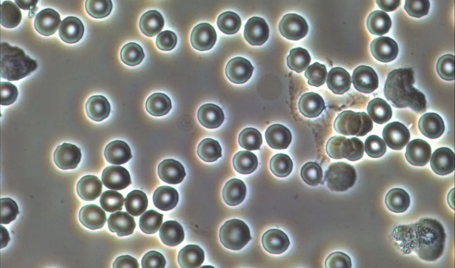
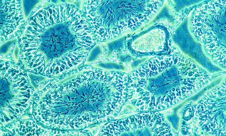
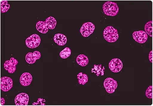
For microscopy techniques, specimen formation is fundamental. Because of the ideal biological specimen, the microscopy technique can be easily used. There are many good reasons to examine the biological cells and tissues under an inverted microscope or simple microscope. The medical and biological research is underpinned by the internal structural details of the cells and tissues. It is seen that in the normal healthy state, all cells and other tissue elements are in good regular shape and recognizable patterns. But in the diseased forms, all such patterns get irregular due to the presence of chemical and physical influences of other elements. This is diagnosed with the help of microscopic techniques like phase contrast microscopy, inverted microscopy, darkfield microscopy, brightfield microscopy, etc.
The detection of diseases becomes easy with an ideal specimen formation. Identification of all these ailments is based on the histopathology and cytopathology of the cells and tissues. This marks the ways for the specializations of modern medicine. In this method, microscopy techniques play an important role in pathological as well as biological research. The study of parasites like viruses, bacteria, etc is detected by the microscopy techniques like phase-contrast microscopy. For the ideal microscopic detection, biological specimens are required. With the help of skilled persons, the formation of biological specimens is done which is fundamental to the microscopy technique.
All types of biological specimens are visualized under the cell culture microscope, culture microscope, and tissue culture microscope. In the scientific world, many types of microscopes are used to observe cells and tissues. The most employed form of microscopy technique is as follows :
These microscopy techniques are used to illuminate the biological specimens with a beam of light rays that pass through a microscope trinocular head. The same happens with the electron microscope, but with a beam of electrons.
In the following ways, the cells and the elements are preserved for the microscopic analysis :
For analyzing the various biological specimens, one requires the ideal biological specimen and that requires the good procedure of biological specimen.
Following are the steps are required for the formation of biological specimens :
It is seen that all fresh tissues are very delicate and easily distorted and can get easily damaged. Due to this fragility of tissues, they can be cut into thin sections of slices. For transforming the cut section into biological specimens, the fixatives are used which helps in the process of preservation of cells and tissues. For fixation, it is broadly divided into the following types :
It is seen that many different biological specimens come from several different sources. This ranges from very large specimens to tiny sources. The fragments are cut down to their cellular or tissue level. Following are the sources of biological specimens used in histopathology systems or biological research :
Fixation of a biological specimen is a crucial step in the preparation of specimens for the microscopy technique. The main objective of fixation is to prevent the cells and tissues from decaying. It helps to preserve the cells and tissues in a ‘life-like state. This is done by stopping the enzyme killing and microorganisms killing and hardening of sections of cells and tissues. All this is done by maintaining the molecular structure intact within cells and tissues.
As soon as fixation is initiated, the separation of the specimen from its blood supply is done. This ensures a good result for the biological specimens. For the fixation process, the most common fixative agent is formaldehyde that is used in the form of a phosphate-buffered solution, called formalin. Usually, all biological specimens are used for analyses under an inverted microscope or simple microscope, the samples are immersed in formalin for 6 to 12 hours, before the process.
Grossing refers to a cut-up of the cells and tissues which involves careful examination and description of the biological specimen. This also includes appearance, dimensions, and several pieces. In the case of larger biological specimens, it requires further dissection to produce representative pieces from the concerned areas. On the contrary, in the case of small biological specimens, no further cutting is required. They can be further processed.
Conclusion
The best trinocular microscope is used to examine the biological specimens for better examination. For better observational study and biological research, specimen formation is mandatory which should be done carefully.