Sample Preparation
Homogenization
Heating and Mixing
Electrophoresis and Blotting
Polyacrylamide Gel Electrophoresis
Agarose Gel Electrophoresis
Western Blotting
Power Supplies
PCR & qPCR Thermal Cycler
Thermal Cycler (PCR)
Real-time Thermal Cycler (qPCR)
PCR Workstations & Cabinets
UVP BioImaging Systems
Molecular Spectroscopy
Lab Equipment
Ultraviolet Products
Hybridization Ovens
UVP Incubator
UV Crosslinkers
UVP Benchtop Transilluminators
Thermal Mixers
Electrophoresis & Blotting
Thermostats
View All
Fume hood
Laminar Airflow
Biosafety Cabinet
Autoclave
Centrifuge
pH Meter
Shaker & Mixer
Orbital Shaking Incubator
BOD Incubator
Heating Oven
Water Purification System
Aermax - Air Purification
Medical Oxygen Concetrators
Hygiene Solution
-150°C Cryogenic Freezer
-86°C Ultra Low Temp Freezer
-40°C Low Temp Freezer
-18 ~ -25°C Biomedical Freezer
-20°C Biomedical Freezer
4° ± 1°C Blood Bank Refrigerators
2~8°C Pharma Refrigerators
2~8°C ICE Lined Refrigerators
-25°C ~ + 4°C Mobile Freezer/Collers
20~24°C Blood Platelet Incubators
Ice Machines
Coldrooms
Mortuary Chambers
All microscopy techniques are used to magnify biological specimens in labeled and unlabeled states. It helps in visualizing structures of various science and biological specimens with detailed information. This information is good for making observations for a concerned biological specimen. For a researcher or scientist, choosing the right microscopy technique for the magnification is a prerequisite thing for an experiment. Many microscopic techniques like phase contrast microscopy, inverted phase microscopy, brightfield microscopy, widefield microscopy, darkfield microscopy, etc, are used to visualize the thick and thin biological specimens.
The fluorescence microscopy techniques are also useful in magnifying the various biological specimens under the trinocular head through the help of fluorescence light lamps. In this technique, the fluorescent dyes and proteins are used to label the concerned biological specimen for the magnification process. This makes the detection process easier for the concerned biological specimen. The main purpose of any microscopy technique is to fully understand the biological sample in terms of its structure, components, and various other aspects. Some of the microscopy techniques give out 3D images of the biological sample which helps in making meaningful observations about it. Thick biological specimens are somewhat harder to visualize than the thin specimens because of the additional layers of structure. But with an efficient microscopy technique, one can visualize thick biological specimens also with ease.
To create a perfect 3D image of a biological structure, one must consider the optics nature used in the microscopy technique. The optics of the light used in the concerned microscopy technique, have a limited field of view and depth of the concerned field used. So, the signals traveled along the path of light create a field over the surfaces of the biological specimens to create their 3D images. These signals are formed in areas both in focus and areas out-of-focus to form the meaningful images of the concerned specimen. In addition to these, diffraction and refraction processes also play an important role in the formation of 3D images. But when these processes of light do not play their distinct roles, the images got fuzzy and no insights can be formed with them.
In historic terms, it is said that the deconvolution techniques are used to eliminate out-of-focus light from the microscopy light. With new and good advancements in microscopy techniques, now to eliminate these errors, computational techniques are used. These systems help to generate the 3D images with full efficiency and proficiency. Within this system, all 3D structures are widely visible to researchers, so that the structures can be inferred.
Every microscopy technique user wants clear and detailed information on the structure of the specimens. It is the most wanted feature of any lab fellow or researcher. But despite using the most efficient microscopy technique, still, images get blurry. This is because the microscopy technique used has the limited capability to reflect the optical light path. This hinders the formation of 3D images. All light sources used in a fluorescent microscopy technique use the scattered lights to form the images. In an inverted phase contrast microscope, the light paths are diffracted in such a way that it forms the phase differences that help in forming 3D images. To visualize the thick biological specimen, fluorescence microscopy techniques are used with elegance.
Still, when the optical light path gets obstructed by other means, the images are called blurry. The scattered lights meet up over the biological specimen, all get obstructed with differences of refractive indexes. This makes the images blurry. To overcome the problem of blurry images of the thick specimen, different approaches have been developed in the form of microscopy techniques. The advancements have led to the magnification of thick specimens
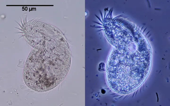
A phase contrast microscopic device tends to manipulate the light paths through the use of strategically placed rings. This is done to illuminate transparent samples and thick segments as well. Fritz Zernike developed the phase contrast microscopy technique in the 1930s. In this microscopy technique, the parallel beams of light are made to pass through the biological specimen of different densities. These microscopes are also called the culture microscope, cell culture microscope, and tissue culture microscope. When phase contrast microscopy technique is combined with inverted microscopy technique. It is called an inverted phase microscope and an inverted phase contrast microscope.
An inverted phase microscope contains special condensers that throw light “out of phase”. All the interferences in the wave nature of light occur in this microscopy technique. Here, the phase rings cause it to pass through the object at different speeds. All Internal details of organelles of live, unstained organisms (e.g. mitochondria, lysosomes, Golgi body) can be seen clearly with phase-contrast devices. Even the best trinocular microscope also has the feature of interference to cause the generation of phase contrasted images and form the sharper images of the thick specimen.
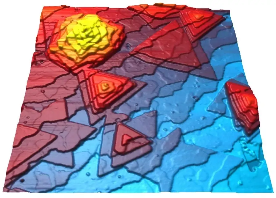
In biology and research labs, the dark field microscope is used for the illumination of unstained thick biological samples. The microscopy technique helps the 3D images to appear bright and fully contrasted images in the darker background. This dark background helps in the perfect illumination of all thick biological specimens. All obtained 3D images are full contrasted like images of phase contrast microscopy. Darkfield microscopes can be also used with electron as well as light microscopy techniques. It has the feature of magnifying the small molecule to a larger resolution of pixels. A camera for a trinocular microscope is fitted which can also be attached with the dark field microscope. It can help in recording the image of specimens. In this mode, no staining of the biological specimen is done. The darkfield microscope illuminates the images against the darker background.
The dark field microscopy technique generates images in their sub-resolution features that enable the perfect magnification and hence, results in sharper images. The high angle scattering of light rays occurs in this microscopy technique. Darkfield microscopy is used for both unstained and stained objects. All thick biological specimens are also get illuminated in the darkfield microscopy technique.
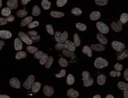
This type of microscopy technique is used for forming images of the thick biological specimens by using fluorescence probes. Staining with fluorescent dyes or proteins is done to form the sharper images under the inverted microscope of fluorescent nature This microscopy technique is used widely to gain information about the structures of the biological specimens. The light source can be electron beams or simple light rays. Here, the microscopy technique is used to illuminate the images with more fluorescent capability than other microscopy techniques. The transmission electron and scanning probe microscopy techniques are the types of fluorescence that help in studying the structures of thick biological specimens.
Conclusion
The microscopy techniques are employed in a biology lab, to gain information about the biological specimens. All techniques are different in their way of action and working. One thing that remains the same is the purpose of visualizing the thicker biological specimens. One can check the trinocular microscope price online as well as offline mediums. All lights get scattered and then culminates over the surface of biological specimens, to form sharper images.