Sample Preparation
Homogenization
Heating and Mixing
Electrophoresis and Blotting
Polyacrylamide Gel Electrophoresis
Agarose Gel Electrophoresis
Western Blotting
Power Supplies
PCR & qPCR Thermal Cycler
Thermal Cycler (PCR)
Real-time Thermal Cycler (qPCR)
PCR Workstations & Cabinets
UVP BioImaging Systems
Molecular Spectroscopy
Lab Equipment
Ultraviolet Products
Hybridization Ovens
UVP Incubator
UV Crosslinkers
UVP Benchtop Transilluminators
Thermal Mixers
Electrophoresis & Blotting
Thermostats
View All
Fume hood
Laminar Airflow
Biosafety Cabinet
Autoclave
Centrifuge
pH Meter
Shaker & Mixer
Orbital Shaking Incubator
BOD Incubator
Heating Oven
Water Purification System
Aermax - Air Purification
Medical Oxygen Concetrators
Hygiene Solution
-150°C Cryogenic Freezer
-86°C Ultra Low Temp Freezer
-40°C Low Temp Freezer
-18 ~ -25°C Biomedical Freezer
-20°C Biomedical Freezer
4° ± 1°C Blood Bank Refrigerators
2~8°C Pharma Refrigerators
2~8°C ICE Lined Refrigerators
-25°C ~ + 4°C Mobile Freezer/Collers
20~24°C Blood Platelet Incubators
Ice Machines
Coldrooms
Mortuary Chambers
Microscopes are a crucial part of any research laboratory. With the help of a microscope, any type of observation can be done on cell cultures and tissues. An inverted microscope is somewhat similar to a simple, traditional microscope. It has the same principle of magnification as the conventional microscopes used in the lab. The only difference between the two microscopy techniques is the nature of the inverted stage in the inverted phase microscope. More or less the components are also the same in the inverted microscope. But the components are all placed in an inverted manner, giving the name upright and inverted microscope.
An inverted microscope comes up with its light source and a set of condenser lenses. These components make the magnification of biological samples easy and subtle. The magnification components are located at the top of the microscope, pointing in a downward direction. On the other side, the objectives and turrets are placed below the stage pointing in an upward direction. The stage used here is stationary in position and does not move at all. However, researchers and scientists can adjust their focus through the condenser lens fitted to the inverted phase-contrast microscope. The biological specimens are observed from the downward fashion. For example, when you observe a part of the tissue in an inverted microscope, you will observe it from its bottom region to the upward region. When the inverted microscopy technique is coupled with the phase-contrast microscopy techniques, it results in the inverted phase-contrast microscope. The resultant images of this type of microscope are highly contrasted and well detailed.
The microscope’s invention dates back to 1850, where the faculty member of Tulane University worked hard for inventing the device. Lawrence Smit, with his friends, invented the first inverted microscope which can be used for the observation of biological specimens in a laboratory. Later, an esteemed company began to manufacture the inverted microscopes on a large scale. From its invention to till now, the inverted microscope has built its popularity base in the scientific world. These microscopes give a lot better magnification than conventional ones. The range is 4x to 40x magnification which is an ideal magnification value for all biological specimens. Due to this feature of an inverted trinocular compound microscope, it is also known as cell culture microscope, culture microscope, and tissue culture microscope.
The inverted phase microscope comes with three to six objective lens that helps in magnifying the images of biological molecules. A condenser lens is also fitted to the trinocular head of the microscope which helps in observations. Live-cell imaging can also be possible with the inverted phase microscope. All living cells are observed through the bottom of the culture vessel. Because of its magnifying feature, the best trinocular microscope is highly famous among researchers and scientists in the laboratory. With this, it also has mere advantages over upright and normal microscopy techniques.
The principle of an inverted microscope is the same as that of a normal microscopy technique. The upright microscope also follows the same principle of magnification. But there are some differences in the case of inverted microscopes. Unlike the normal upright microscopes, the inverted microscope has a condenser lens located on the top of the stage in the microscope. In an actual way, the inverted microscope also has the light source in the top position.
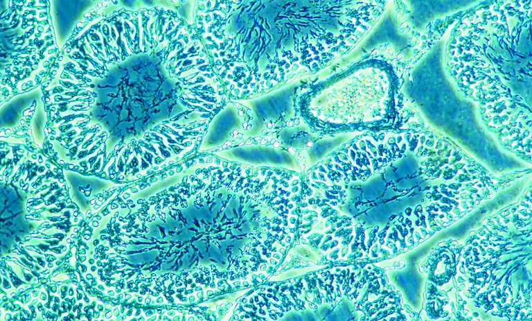
The only principle that governs the formation of images of biological samples is the reflection of light through the condenser lens. The diffraction process also happens in the inverted microscope which helps in the process of magnification. The condenser lens at the top of the device focuses the coming light to the specimen. Light rays reach the biological specimen where it starts reflecting on the surface of molecules. All objective lenses that are located below the stage mediate the light rays through its lens. Here, the objective lens is normally three to six in number. In the inverted phase-contrast microscope, cell culture and tissue cultures are observed with utmost details and structures through the culture vessels. To obtain a good image of the biological specimen, the vessel should be of high optical capacity. So, light rays can be reflected and transmitted. A high-quality coverslip should be used for the observations of live cells and tissues.
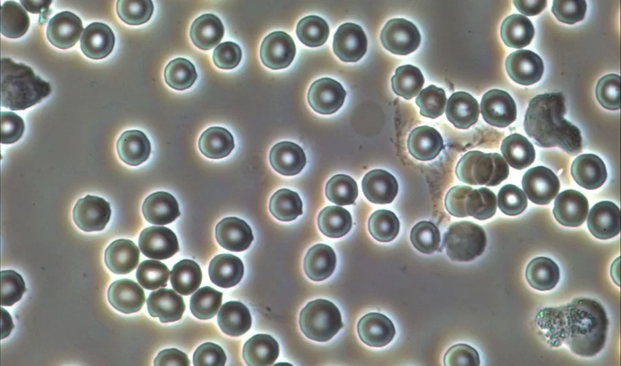
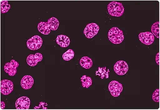
It is seen that the inverted microscopy technique has many applications in the diagnosis and clinical research area. The technique is famous among researchers and scientists due to its magnifying power through the culture vessels. Usually, the cells sink to the bottom of the culture vessel, if no suspension is there. In this case, the observation can be hindered and unclear. The inverted microscope comes to the rescue for this. As in the inverted microscopy technique, the images are obtained from the bottom so the cells that cling to the bottom get magnified. Like this, many applications prevail in the scientific world for inverted phase microscopes.
Following are the applications of inverted microscope that are widely used :
Following are the various benefits of inverted technique :
Conclusion
Due to the inverted microscopy magnifying power, it is more suitable for observing live cells in Petri dishes or cell cultures. The principle of inverted microscopy is the same as the normal, simple microscope. The inverted microscopes come a little more expensive than the simple microscopes. You can check the inverted microscope price online as an offline medium.