Sample Preparation
Homogenization
Heating and Mixing
Electrophoresis and Blotting
Polyacrylamide Gel Electrophoresis
Agarose Gel Electrophoresis
Western Blotting
Power Supplies
PCR & qPCR Thermal Cycler
Thermal Cycler (PCR)
Real-time Thermal Cycler (qPCR)
PCR Workstations & Cabinets
UVP BioImaging Systems
Molecular Spectroscopy
Lab Equipment
Ultraviolet Products
Hybridization Ovens
UVP Incubator
UV Crosslinkers
UVP Benchtop Transilluminators
Thermal Mixers
Electrophoresis & Blotting
Thermostats
View All
Fume hood
Laminar Airflow
Biosafety Cabinet
Autoclave
Centrifuge
pH Meter
Shaker & Mixer
Orbital Shaking Incubator
BOD Incubator
Heating Oven
Water Purification System
Aermax - Air Purification
Medical Oxygen Concetrators
Hygiene Solution
-150°C Cryogenic Freezer
-86°C Ultra Low Temp Freezer
-40°C Low Temp Freezer
-18 ~ -25°C Biomedical Freezer
-20°C Biomedical Freezer
4° ± 1°C Blood Bank Refrigerators
2~8°C Pharma Refrigerators
2~8°C ICE Lined Refrigerators
-25°C ~ + 4°C Mobile Freezer/Collers
20~24°C Blood Platelet Incubators
Ice Machines
Coldrooms
Mortuary Chambers
Microscopy techniques are the backbone of biological research that enables researchers and scientists to evaluate the structures of specimens. With the advent of technology, microscopic techniques have evolved so much. From the first discovered traditional microscope to the high advanced phase contrast microscopy device. The evaluation through the trinocular head of the microscope has changed. One such advancement is the addition of digital devices to the trinocular compound microscope which helps in the perfect evaluation of the biological specimens. When the phase contrast microscopy is combined with the digital device, then the whole technique is called “Digital Pathology”.
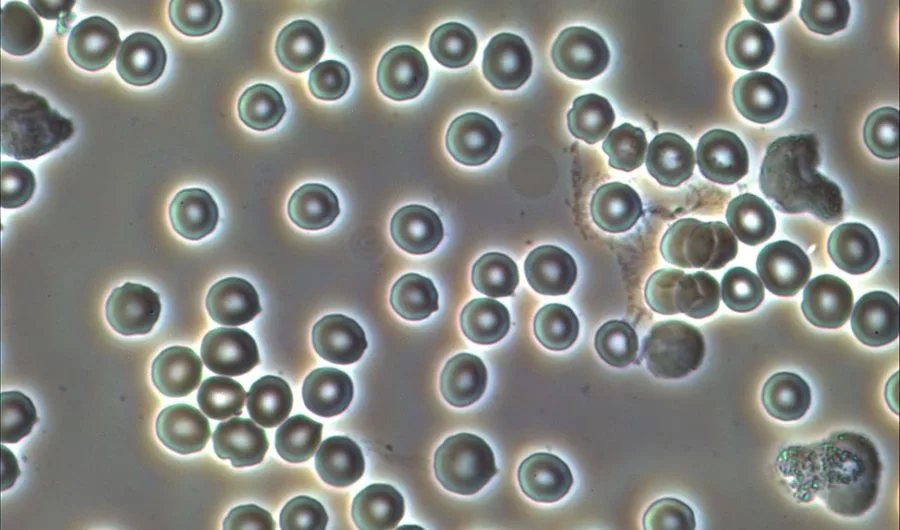
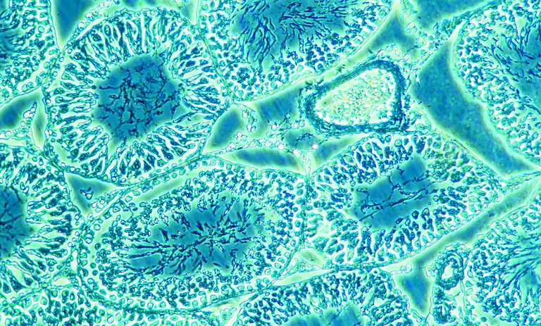
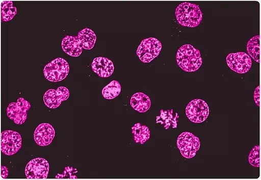
Digital pathology basically includes the acquisition of biological specimens, management, and sharing and interpretation of pathological information. The information includes the slides and data sets inferred from the various biological specimens. All this work is done on a digital device, like laptops or desktops in a digital environment. In this process, the digital slides are formed on the screens of desktops when the glass slides get captured by a scanning device. The scanning microscopes are employed for the process of scanning biological specimens. In this scanning process, the images formed in the computer screens are of high resolution and very structured. The images can also be viewed in a connected mobile screen also. When the microscopes are employed for the evaluation of cell culture and tissue culture, it is called a cell culture microscope, culture microscope, and tissue culture microscope.
To evaluate biological images in a structured way, digital devices are used. The digital screen evaluation incorporates screening, management, and inferring the digital information from the samples. The images formed on the screen are in high resolution, so that perfect evaluation can be done. For scanning purposes, the lab fellows, researchers, and scientists utilize the high-throughput automated digital pathology scanners that enable the whole of the entire glass of the inverted phase microscope. Usually in digital pathology techniques, brightfield, darkfield, and phase contrast microscopy techniques are used for generating good images. However, fluorescence microscopes are also used in this manner.
All digital slides created in this process are made sharable to a network of people by specialized digital pathology software among the researchers. This is marking the characteristic of digital pathology. Moreover, the automated image analysis devices can also be utilized to assist in the interpretation and quantification of biomarker expression within tissue specimens. Various cell and tissue culture samples are marked easily with the image analysis tool.
History Of Digital Pathology Or Screen Evaluation
The history of digital pathology dates back over 100 years. It has been in the scientific world for such a long period. In the initial days, digital pathology was carried out with the help of specialized equipment to capture images from the inverted phase contrast microscope. From the microscopic views, the images were transformed into photographic plates. And the technique of sharing the photographic plates with a group of people has been around in biological research for 50 years. Today, it has evolved so much with the advent of technology which makes it more advantageous than the traditional methods of screening biological specimens using an electronic device.
The whole objective of digital screen evaluation is to enable the pathologists, researchers and scientists to evaluate, engage and collaborate rapidly and remotely with a lot of transparency and consistency with biological specimens’ results. The outcome of all these results in the efficiency and productivity of the biological research process. This has also enhanced translational research, computer-aided diagnosis, and personalized medicine.
Steps To Digital Screening Evaluation And Pathology
The following are the steps to successfully evaluate the biological specimen’s images under an inverted microscope.
Pathology starts with the best trinocular microscope and the collected tissue specimen. The scans which result from the digital pathology process are called digital scans. A lot more has changed in the digital screening evaluation technique with the pace of technology. With this, the pathology technique has gained some advantages which are considered by the researchers and scientists as boons in the evaluation of specimens.
Following are the benefits of the digital pathology or screening evaluation :
Conclusion
In the long run, digital pathology ensures greater access to tissue and slide-based material outside of the laboratory. The images formed on the computer screen are in high resolution and also mark the cells and tissues. Users of digital pathology can also have access to all their digital courses or processes through the internet or standard web browser. Nowadays, the digital pathology or screening process is done in the laboratory to extract good results out of the specimens. Moreover, the data sets can be transferred among people in a remote way with digital pathology or screening process.