Sample Preparation
Homogenization
Heating and Mixing
Electrophoresis and Blotting
Polyacrylamide Gel Electrophoresis
Agarose Gel Electrophoresis
Western Blotting
Power Supplies
PCR & qPCR Thermal Cycler
Thermal Cycler (PCR)
Real-time Thermal Cycler (qPCR)
PCR Workstations & Cabinets
UVP BioImaging Systems
Molecular Spectroscopy
Lab Equipment
Ultraviolet Products
Hybridization Ovens
UVP Incubator
UV Crosslinkers
UVP Benchtop Transilluminators
Thermal Mixers
Electrophoresis & Blotting
Thermostats
View All
Fume hood
Laminar Airflow
Biosafety Cabinet
Autoclave
Centrifuge
pH Meter
Shaker & Mixer
Orbital Shaking Incubator
BOD Incubator
Heating Oven
Water Purification System
Aermax - Air Purification
Medical Oxygen Concetrators
Hygiene Solution
-150°C Cryogenic Freezer
-86°C Ultra Low Temp Freezer
-40°C Low Temp Freezer
-18 ~ -25°C Biomedical Freezer
-20°C Biomedical Freezer
4° ± 1°C Blood Bank Refrigerators
2~8°C Pharma Refrigerators
2~8°C ICE Lined Refrigerators
-25°C ~ + 4°C Mobile Freezer/Collers
20~24°C Blood Platelet Incubators
Ice Machines
Coldrooms
Mortuary Chambers
It is said that in a biological research lab, there are a lot of instruments that help in retrieving the information from reaction samples. One such process is the instrument called spectrophotometry which is mainly concerned with the qualitative measurements done in the lab. It is one of the branches of electromagnetic spectroscopy that helps in measuring the details of reflection and transmissions of light beams. Here, the transmission is the property of the light beam which is meant to pass through the device. A double beam spectrophotometer consists of photometers that radiate the light beams for the measurement process. These photometers are called spectrophotometers that can measure the intensity of light beams throughout the device. The light beams are recorded with different wavelengths within the single and double beam spectrophotometer.
A double beam spectrophotometer usually uses ultraviolet rays, infrared rays, and visible rays for the transmission process. However, the modern and recent spectrophotometers use a wide spectrum of electromagnetic wavelengths, that includes ultraviolet rays, x-rays, visible rays, infrared rays, microwave rays for the reflection and transmission process.
The main work of a double beam spectrophotometer is to determine the amount of light for a specific wavelength. Here, the beams of light are absorbed by the analyte within a biological sample. The specimens or samples in spectrophotometry can be gases, liquids. The analytes are dissolved in a solvent in the process of spectrophotometry. For the solvents used here, the solid pellets are mixed with a transparent matrix for being used as an analyte for spectrophotometry. The disks for the double beam spectrophotometer are made by using a pellet press which has a disk suspended in the biological samples through which the light beams pass on.
A double beam spectrophotometer is usually used for the measurement of transmittance or reflectance of analytes in a sample. The samples used here are transparent or opaque solids, like polished glass, or gases that can be used as solvents. It is seen that many types of biochemicals come colored, due to which they absorb visible light or ultraviolet light. The transmitted light can be measured by colorimetric procedures within the spectrophotometer. Even the colorless biochemicals used here can be converted to colored compounds for the process. It is suitable for chromogenic color-forming reactions to result in compounds suitable for colorimetric analysis in a single and double beam spectrophotometer.
Though it is seen that they can also be designed to measure the diffusivity of many analytes. This could happen on any of the listed light ranges which any researcher uses in his or her biological research. Here, the light rays usually cover around the range of 200 nm - 2500 nm by using different controls and calibrations used in the spectrophotometers. Within these ranges of light beams, calibrations are needed on the instrument using standards that vary in type depending on the wavelengths of light in the photometric determination.
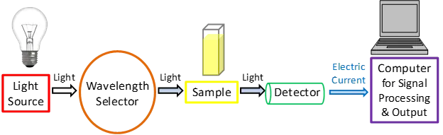
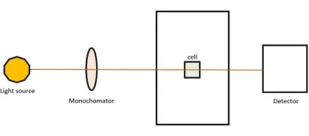
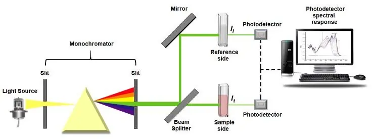
The main driving principle for double beam spectrophotometer is the reflection and transmittance of the light rays. In any single and double beam spectrophotometer, the main element is the light source which has strikes with strong light beams. These light beams depend on the wavelength of interest which are electrically powered ultraviolet and visible rays or infrared rays. Here, the monochromator selects the light beams of a specific analytical wavelength from the light source. The wavelengths are chosen from the spectrums of the light rays passing through the double beam spectrophotometer. Light ray’s analytical wavelength is selected based on the absorbance characteristics of the analyte used in the instrument. Monochromators are instruments whose main purpose is to allow polychromatic light beams to enter into the entrance slit of the monochromator instrument. It also allows a single light ray wavelength, which is monochromatic, to go out via the exit slit through the device. This exciting, narrowly defined light beam consists of a small region of the electromagnetic spectrum of light. The spread and the electromagnetic band-pass of the wavelengths depend on the slit methods or openings of the monochromator attached with the spectrophotometer. These monochromators are adjustable for manipulating the light rays in the single and double beam spectrophotometer. Here, the quality of the light rays which disperses elements in the monochromator helps in the transmission and reflection process.
In the instrumental design of a double beam, a spectrophotometer has the light source from which the light beams strike the samples. The light rays are diverted by right angles within the spectrometry process. This happens by rotating a disk with three distinct panels in the spectrophotometers. One of the sectors of the device allows the light beam to pass straight through the disk without striking. And then another point consists of a mirror surface, with a third point which is black. When the light beam passes through the disk panels, it shines directly into the specimen cells. If the specimen is a liquid or gas, then this cell sample contains a cuvette and is made of transparent stuff that does not absorb light rays in the spectral region of interest of the double beam spectrophotometer. The analyte within the sample is dissolved in a solvent held in the cuvette part of the double beam spectrophotometer. When the source light rays are reflected at right angle degrees by the rotating disk, instead of striking the biological specimen. The light rays pass through a cuvette part in the reference cell which contains only the solvent. This all helps in transmitting the light rays and the process of reflection.
During the third sequence of light rays in the spectrophotometer, when the black sector part blocks the light rays source beam and no light passes through the disk to the sample. As a result, no light rays arrive at the photo transducer in the instrument. This part of the cycle of light rays is used by the computer to quantify and measure the dark current. This dark current is defined as the amount of light produced by the photo transducer circuit in a double beam spectrophotometer. This happens when no light rays strike on the photo transducer part. The dark current can be reduced from the overall light measurements made by the spectrophotometer.
After traveling through every part of the specimen cell or sample cell the light rays which do not get absorbed, most of them are directed onto the photo transducer or light detector in the spectrophotometer. This part of the component converts the arrival of photons in the light rays into an electrical signal. This conversion is made through the computer screen. In this way, the light paths through the spectrophotometer need not be in a straight line. It can be disarrayed also. As it is seen that the light beam of rays can be redirected using mirrors in the device and can also be scattered through the help of collimation.
Conclusion
The double beam spectrophotometer helps transmit the light rays which are after the result of the reflection process. The application of a single-beam spectrophotometer is the same as the double beam spectrophotometer. The light source is used for striking the light beams on the biological specimens and then digitizing the results using a computer system.