Sample Preparation
Homogenization
Heating and Mixing
Electrophoresis and Blotting
Polyacrylamide Gel Electrophoresis
Agarose Gel Electrophoresis
Western Blotting
Power Supplies
PCR & qPCR Thermal Cycler
Thermal Cycler (PCR)
Real-time Thermal Cycler (qPCR)
PCR Workstations & Cabinets
UVP BioImaging Systems
Molecular Spectroscopy
Lab Equipment
Ultraviolet Products
Hybridization Ovens
UVP Incubator
UV Crosslinkers
UVP Benchtop Transilluminators
Thermal Mixers
Electrophoresis & Blotting
Thermostats
View All
Fume hood
Laminar Airflow
Biosafety Cabinet
Autoclave
Centrifuge
pH Meter
Shaker & Mixer
Orbital Shaking Incubator
BOD Incubator
Heating Oven
Water Purification System
Aermax - Air Purification
Medical Oxygen Concetrators
Hygiene Solution
-150°C Cryogenic Freezer
-86°C Ultra Low Temp Freezer
-40°C Low Temp Freezer
-18 ~ -25°C Biomedical Freezer
-20°C Biomedical Freezer
4° ± 1°C Blood Bank Refrigerators
2~8°C Pharma Refrigerators
2~8°C ICE Lined Refrigerators
-25°C ~ + 4°C Mobile Freezer/Collers
20~24°C Blood Platelet Incubators
Ice Machines
Coldrooms
Mortuary Chambers
UV-Vis spectrophotometry is a laboratory technique used in the measurement of absorbance of light across ultraviolet and visible regions. When the incident light strikes the sample, it is usually reflected, transmitted, or absorbed. The absorbance of light causes the transition of molecules from the ground state to the excited state. UV-Vis spectrophotometer, an analytical instrument, serves this principle and measures the intensity of light absorbed by the molecule.
To obtain high precision and a more dynamic range, it is necessary to understand the design of a UV-Vis spectrophotometer. Further, to disperse the wavelengths of light and detect the absorbed wavelength in the sample, it is essential to know about the working of the UV-Vis spectrophotometer. This article details the way of designing a cheaper and efficient UV-Vis spectrophotometer and the way of working a UV-Vis spectrophotometer.
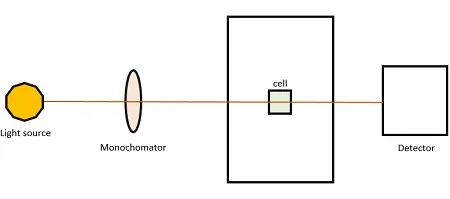
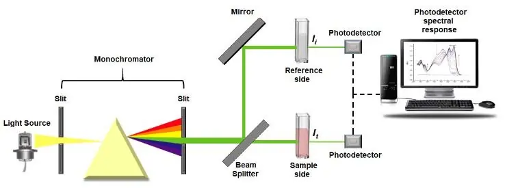
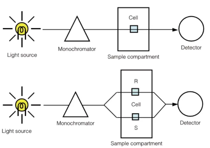
The design of the UV-Vis spectrophotometer includes both the parts of the spectrometer and photometer. The spectrometer part contains a light source, sample cell, monochromator, and slits. The microcontroller controls grating in the monochromator, whereas the photometer part consists of a photodiode, preamplifier, and data acquisition system.
There are many parts in the instrumentation of the UV-Vis spectroscopy system, which are indispensable in the functioning of the UV-Vis spectrophotometer. Ultraviolet-visible spectrophotometer system focuses electromagnetic radiation from the light source to the sample. Depending on the configuration set in the system, light is transmitted through the sample or reflected off it. Then, the light is collected from the sample through reading.
Initially, light is focussed into the entrance slit of the monochromator from the source. Monochromator uses dispersing elements, namely optical grating to separate the light by wavelength. The light is passed into a charged coupled device (CCD), which is made up of individual tiny detectors, hence the intensity of light at each wavelength will be measured. CCD is read-off to a computer and the result obtained is a spectrum, which shows the intensity of each wavelength of light. Spectrophotometers are able to measure the electromagnetic radiation from ultraviolet to infrared. Spectrum will show the intensity of light versus the wavelength.
There are 3 classifications of UV-Vis spectrophotometer. They are as follows:
In a single-beam spectrophotometer, light from the monochromator alternatively passes through reference and sample solution. As it is affected by power supply fluctuations, this type is not suitable for high-demanding pharmaceutical and quality inspection experiments.
In a double-beam spectrophotometer, there are two wavelengths of monochromatic light, which alternatively irradiate samples at regular intervals. It performs better for turbid samples, eliminating the light source instability and detector sensitivity changes.
In a split beam spectrophotometer, the light beam from the monochromator is split into two beams. One beam passes through the detector directly and the other one passes through the sample and reaches the detector. It is able to monitor errors in the light source, but can not eliminate the effects in the reference cell.
Conclusion
Design of a spectrophotometer should be done with a low cost, good quality and low power consuming materials. The materials used should be sensitive to minute changes and accurate, ensuring good reproducibility. In addition, deviation should be minimal between already established equipment and newly designed instruments.It should be made sure that the designed equipment will be compatible to all types of samples.