Sample Preparation
Homogenization
Heating and Mixing
Electrophoresis and Blotting
Polyacrylamide Gel Electrophoresis
Agarose Gel Electrophoresis
Western Blotting
Power Supplies
PCR & qPCR Thermal Cycler
Thermal Cycler (PCR)
Real-time Thermal Cycler (qPCR)
PCR Workstations & Cabinets
UVP BioImaging Systems
Molecular Spectroscopy
Lab Equipment
Ultraviolet Products
Hybridization Ovens
UVP Incubator
UV Crosslinkers
UVP Benchtop Transilluminators
Thermal Mixers
Electrophoresis & Blotting
Thermostats
View All
Fume hood
Laminar Airflow
Biosafety Cabinet
Autoclave
Centrifuge
pH Meter
Shaker & Mixer
Orbital Shaking Incubator
BOD Incubator
Heating Oven
Water Purification System
Aermax - Air Purification
Medical Oxygen Concetrators
Hygiene Solution
-150°C Cryogenic Freezer
-86°C Ultra Low Temp Freezer
-40°C Low Temp Freezer
-18 ~ -25°C Biomedical Freezer
-20°C Biomedical Freezer
4° ± 1°C Blood Bank Refrigerators
2~8°C Pharma Refrigerators
2~8°C ICE Lined Refrigerators
-25°C ~ + 4°C Mobile Freezer/Collers
20~24°C Blood Platelet Incubators
Ice Machines
Coldrooms
Mortuary Chambers
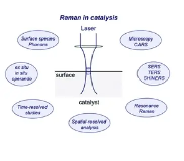
Raman spectroscopy is one of the experimental, sophisticated and spectroscopic techniques, which are used in research and quality control laboratories. Raman spectroscopy imaging, Raman imaging, and Raman shift are the alternative times used for this technique.
This article brings light to the advances of Raman spectroscopy specifically in the areas of cell biology and natural tissues and importance of characteristic peak frequencies. Some of the most widely utilized peak frequencies and their assignments are summarized.
The study aims at the preparation of a database of molecular fingerprints, which aids researchers in the definition of the chemical structure of the biological tissues, through the introduction of most of the significant peaks present in the natural tissues.
Though different methods are applied, there is a considerable similarity in the definition of peaks of the identical areas of the Raman spectrum. As a consequence, the preparation of a unique collection of frequencies encountered in the study of Raman spectroscopy leads to substantial improvement in the quality and quantity in data of Raman spectra and their outcomes.
The article provides a concise database on the important Raman lines in Raman spectroscopy for the researchers, who aim to analyze the natural tissues through Raman shift. This also serves as considerable assistance to the people, who focus on the investigation of cancerous tissues through Raman analysis.
It is a widely utilized method for the characterization of biological tissues. It is a technique of choice for scientists for finding the chemical and structural properties of synthetic and natural tissues.
Using peak frequencies at 1300 cm-1 (CH2 twist), 1440 cm-1 (CH2 bend), 1656 cm-1 (C=C), and 1754 cm-1 (C=O), the lipid components and chemical structures can be evaluated well.
Through the peak frequencies at 1300 cm-1(CH2 bend), 1440 cm-1(amide I), 1656 cm-1(C=C), and 1754 cm-1(C=O), protein contents are investigated well.
Definitions of peak possess a momentous influence on the reliability of the results offered by different spectroscopic techniques. Most of the scientists have predominantly utilized the previously published articles for the definition of the data acquired from the collected Raman spectra. In any case, without a detailed and reliable database, which may cover most of the needed peaks in the spectral range, it will be a laborious task for finding the meanings of various unknown peak intensities.
In biological analyses, a wide range of functional groups and chemical bands are attributed to every single peak observed, finding the appropriate meanings that demonstrate the clinical significance of the technique and attaining results. This serves as one of the most significant steps in finalizing a spectroscopic research work.
Using Raman spectroscopy, various tissues like cervical tissue, bone, cornea, lung, breast, epithelial tissue, mammalian cell culture, cancerous cells, meningioma, tissue processing, raft cultures, saliva, anti-cancer drugs, cancer genes, DNA, individual cells, microbial cells, human living cells, mixed cancer cells, human red blood cells, lymphocytes, human coronary arteries, serum, atherosclerotic plaque, heme protein, liver, oral tissue, brain, skin, gastrointestinal tissue, lung, and breast have been investigated.
Raman spectroscopy can monitor changes, which happen due to the proliferation of the cell. This is performed by the investigation of cultures in non-proliferative and proliferative phases of growth, followed by estimation of relative amounts of biochemical components such as RNA, DNA, proteins, and lipids in nuclei and cells. Reproducible differences are detected and quantified using relative amounts and ratios of biochemical compounds. This information can be significant for Raman analysis and detection of rapidly dividing populations of cancer cells in vivo.
Koljenovic and his team performed a study on normal dura mater and meningioma through Raman microscopy for assessment of the possibility of developing an in vivo Raman method for the guidance of meningioma reactions. Raman maps were constructed of cryosections of meningioma and dura, which were obtained from 20 patients. The comparison of maps with histopathology led to the assignment of Raman spectra to either dura or meningioma. Greater dissimilarities exist between dura and meningioma because of the high collagen content in the dura and increased lipid content of meningioma. Based on linear discriminant analysis of Raman spectra, the classification model for dura and meningioma provided an accuracy of 100%. Raman spectra empower the meningioma to be differentiated from the dura. This makes Raman spectroscopy a viable contender for the guidance of surgical resection of meningioma.
Jalkanen and his team utilized vibrational spectroscopy for studying structures of DNA and proteins. In addition, hydration and binding of biomolecules were studied using a combined experimental and theoretical approach. The systematically studied molecules were mainly peptides and amino acids. The aim was to interpret the experimentally estimated vibrational spectra for the molecules to the greatest probable extent and to figure out the function, structure, and electronic properties of the molecules in the various environments. The application of various spectroscopic methods to environmental and biophysical assays is in expansion and therefore, a deeper understanding of the phenomenon from a rigid theoretical basis is needed.
There is an increase in the number of research groups, who are interested in investing time and funds for Raman spectroscopy. The results are useful for speedy and precise interpretation of data. It will help researchers for easy follow-up and easily understand the significant characteristic peaks in Raman spectra of biological tissues. Considering the type of samples, chemical bonds, and functional groups involved, peak frequencies can be located and appropriate interpretations can be performed with confidence. Peaks can be assigned to different biochemical compounds like lipids, nucleic acids, and proteins, which lead to a better correlation between structural, chemical, and medical aspects of spectroscopy. It provides major assistance to spectroscopists and those working in chemistry, tissue engineering, and biomaterials sciences. However, the database should be periodically and continuously updated, so that it can be useful for different methodologies.
Conclusion
A definition of Raman spectroscopy has been given. The significance of Raman spectroscopy in biology has been listed. The concept of characteristic peak frequencies has been stressed. Then, the regions of investigation under Raman spectroscopy have been shown. In addition, three cases of Raman spectroscopy in biological research have been discussed. Further, the pros of utilizing Raman spectroscopy in biological tissues have been briefed.